
Asto CT Equine Tales: Unveiling Journeys of Recovery with the Equina
In the world of veterinary medicine, every horse owner and their equine companion have a unique story to tell, and their road to recovery is often filled with unexpected twists and turns. In this article, we introduce you to three remarkable cases diagnosed with the Equina Computed Tomography (CT) scanner. These stories provide insights into how the Equina by Asto CT has revolutionized the way equine surgeons approach diagnostics and treatment plans, ultimately improving the chances of a successful recovery. Let us delve into the intriguing journeys of Mabel, Roman, and Chester as they navigate their way through the world of equine healthcare.
HI, MY NAME IS MABEL
Mabel is a 5-year-old Quarter Horse mare who runs barrels. Her owner made the decision to bring her to the clinic due to a traumatic injury that occurred in her stall overnight. She had a severe lameness in her left hind leg, and a wound near her fetlock area.


CLINICAL EXAMINATION
Upon initial examination, clinicians took radiographs which showed the presence of several fracture fragments in her fetlock area and swollen soft tissue of the distal limb. Subsequently she was recommended for a CT scan to further investigate in more detail.
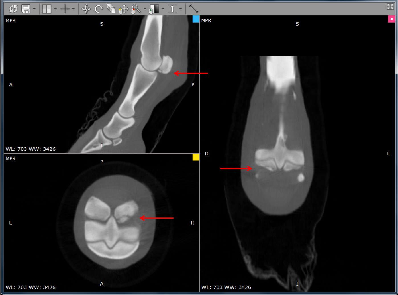
CT FINDINGS - MULTIPLE FRACTURES FRAGMENTS
CT images showed acute, traumatic, complete, comminuted fractures involving the medial proximal sesamoid bone (basilar and abaxial) and third metatarsal bone (sagittal ridge). Additionally, an intracapsular metatarsophalangeal fracture fragment with effusion of the fetlock joint was noted. This condition was challenging to diagnose due to the presence of multiple fractures in the fetlock region.
PROXIMAL SESAMOID FRACTURES
Proximal sesamoid fractures range from simple to complex, and may be articular as well as non-articular. These types of fractures are commonly seen in racehorses due to hyperextension type injuries; however, in this case trauma was the cause of the injury. When a proximal sesamoid fracture occurs, it can result in lameness and discomfort for the horse. They can potentially disrupt the function of the surrounding tendons and ligaments, which are crucial for the horse's movement and support.
SURGERY
Mabel was treated surgically with arthroscopy to remove the fragments and debride the traumatic wound.
WHY IS CT USEFUL?
Asto CT's technology allows veterinarians to obtain three-dimensional images of the horse's limb while the animal remains standing, eliminating the need for anesthesia and providing a more comprehensive view of the injury. This precise imaging was instrumental in diagnosing Mabel’s acute fractures, determining the extent of the damage, and identifying the best treatment plan with minimal complications. With this information, veterinarians can develop surgical and treatment plans individualized to their patient and improve the chances of a successful recovery.
HI, MY NAME IS ROMAN
Roman is a Shire gelding who currently shows at the top-level circuits. His owner made the decision to bring him to the clinic due to a persistent draining tract and emerging complications within his right front foot.


CLINICAL EXAMINATION
Roman was initially treated surgically for quittor, an infection of the collateral cartilage on the lateral side of the foot. Surgery went well, and he went home to continue healing. A couple months later he returned to the clinic for a persistent draining tract. Subsequently he was recommended for a CT scan to further investigate.
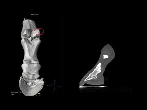
CT FINDINGS - FISTULA WITH SEQUESTRUM
CT images show a draining tract just proximal to the coronary band on the lateral aspect of the foot. Clinicians performed a fistulogram on the horse in the CT suite. They were able to determine the draining tract (fistula) communicated all the way down to the coffin bone where there was a suspected bone sequestrum.
WHAT IS A FISTULA AND SEQUESTRUM?
A fistula is an abnormal tunnel-like tract that connects between the bone sequestrum and the skin in this case. Reason being the body is trying to expel the dead piece of bone with surrounding infection and creates a fistula to do so.
A bone sequestrum in a horse is a piece of dead bone that has separated from the healthy bone. A bone sequestrum that is infected can delay the healing process and it will continue to drain and cause pain until it is removed.
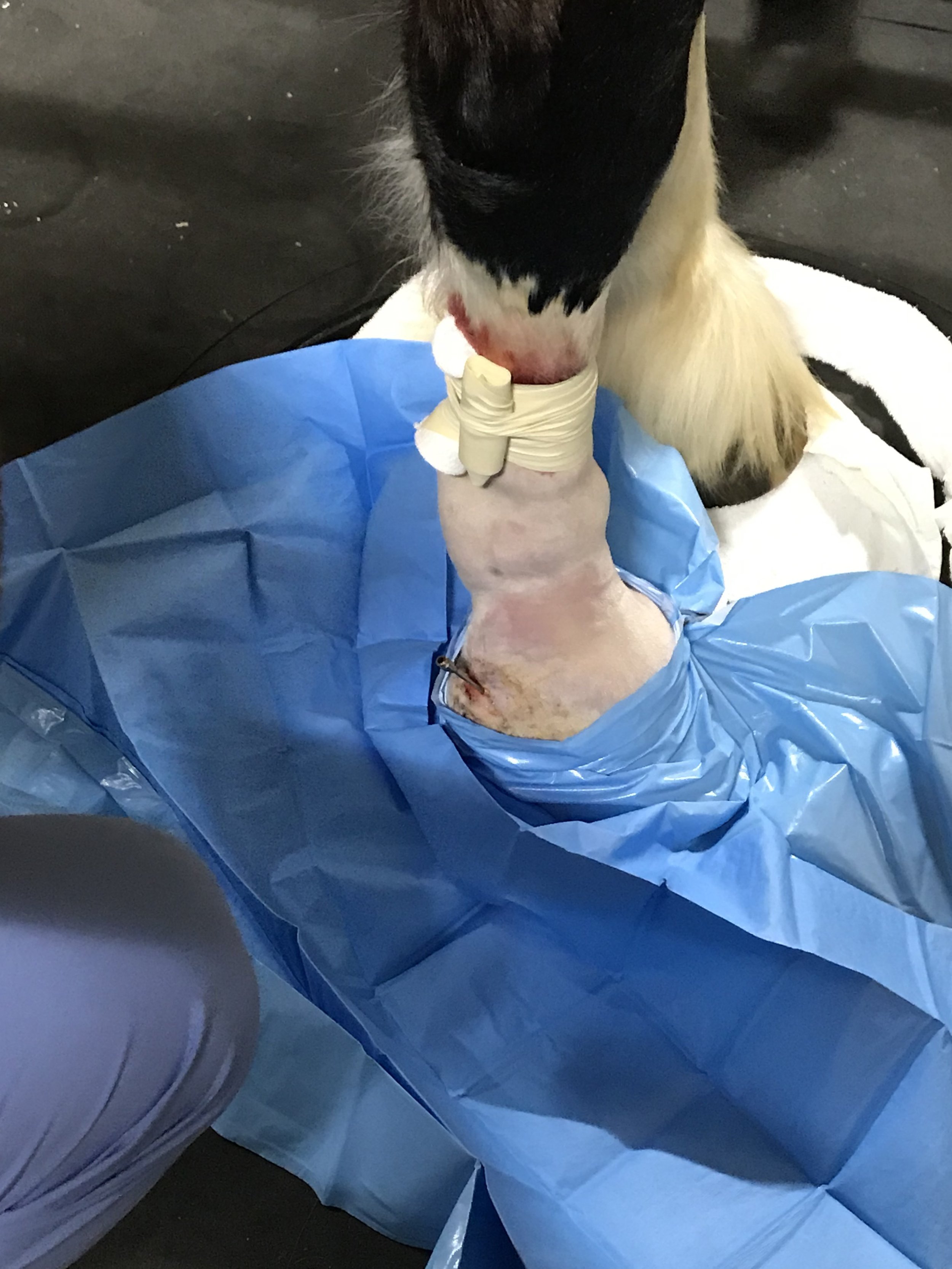
SURGERY
Roman was able to remain standing on top of the CT scanner throughout the entire procedure. The picture shows a cannula that was placed in the draining tract, where contrast was administered. The team successfully debrided the bone sequestrum under CT guidance. Roman is now doing much better at home.
WHY IS CT USEFUL?
Interoperative imaging using the Equina standing CT scanner is particularly useful in cases where standing surgery is an option. CT imaging can be used to create 3D reconstructions of the affected area, which can be incredibly helpful in planning surgical procedures or interventions. These reconstructions allow the veterinarian to assess the sequestrum's relationship to other structures and plan the best approach, minimize damage to healthy tissues, and increase the chances of successful removal.
HI, MY NAME IS CHESTER
Chester is a retired show horse that currently serves as a decorative pasture companion. His owner made the decision to bring him to the clinic due to emerging complications within his nasal cavity.


CLINICAL EXAMINATION
Chester presented to the clinic with nasal discharge and intermittent epistaxis (nosebleed). He was subsequently recommended for a head CT scan in order to further investigate the underlying cause of his symptoms.
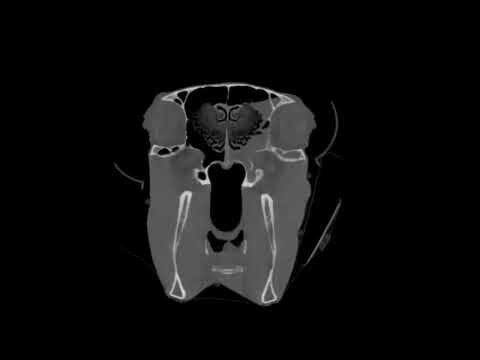
CT FINDINGS - ETHMOID HEMATOMA
CT images show a soft tissue mass in the left nasal cavity. This mass is intermittently associated with the left ethmoid turbinate and surrounded by soft tissue that has fluid attenuation and fills the surrounding sinuses. Right side is normal.
WHAT IS AN ETHMOID HEMATOMA?
An ethmoid hematoma is a well-recognized but poorly understood disease in horses. Affected horses classically exhibit bloody nasal discharge originating from a hematoma-like mass within the ethmoid turbinates of the nasal cavity. source
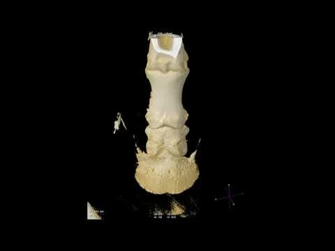
SURGERY
The mass was removed through a frontonasal flap. The 3D reconstruction shows staples and sutures that closed the flap. Chester is reportedly doing well at home. Histopathology – findings consistent with ethmoid hematoma with bacterial colonies.
WHY IS CT USEFUL?
CT is useful in ethmoid hematoma cases where the mass cannot be viewed endoscopically because of the size or radiographically. CT scans provide a three-dimensional view of the hematoma and its relationship with adjacent structures. This helps surgeons plan the most appropriate treatment approach, whether it involves surgical removal or other therapeutic options.
These stories of Mabel, Roman, and Chester emphasize the significance of advanced diagnostic tools like the Equina by Asto CT in equine medicine. These tools have not only demystified complex conditions but have also influenced successful treatment strategies. As we conclude these captivating case studies, we celebrate the remarkable progress in veterinary medicine, ensuring horse owners can pursue their equine companion’s path to recovery with renewed hope, thanks to technology, expertise, and unwavering commitment.