
Occipital Bone Lysis and Hyperostosis
FEATURED WINNER – Limb Category
Occipital Bone Lysis and Hyperostosis
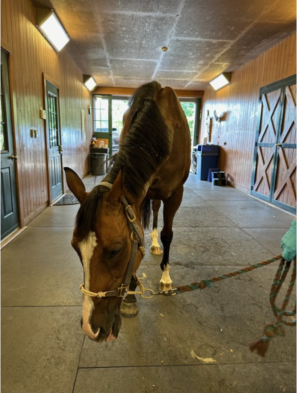
15-year-old Thoroughbred gelding
Virginia Equine Imaging
Dr. Jack Caldwell
Highest Scoring Entry with a score of 33 out of 35 points.
This case was recognized for its exceptional 3D reconstruction quality and its ability to clearly define complex bony pathology in the occipital region—information that would have been difficult to fully appreciate using traditional imaging alone.
Case Summary
A 15-year-old Thoroughbred gelding was referred for full-body nuclear scintigraphy due to bilateral hindlimb lameness, decreased cervical range of motion, and Grade 2–3 ataxia. The gelding had a prior history of guttural pouch mycosis treated three years earlier with coil embolization of the left carotid artery. Nuclear scintigraphy revealed intense focal radiopharmaceutical uptake at the external occipital protuberance (poll) and C1, prompting advanced CT evaluation of the caudal head and cranial cervical spine.
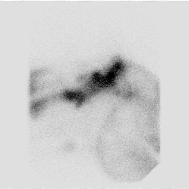
Nuclear Scintigraphy
Clinical Signs
At rest, the gelding stood with his head lowered and deviated to the right.
Diagnosis
CT imaging demonstrated extensive occipital bone lysis and hyperostosis, regional infiltrative hypoattenuating soft tissue causing spinal cord compression, and marked thickening of the left stylohyoid bone and temporohyoid articulation. Findings were most consistent with fungal osteomyelitis and meningitis, given the history of guttural pouch mycosis.


Outcome & Post-Mortem Findings
Due to the poor prognosis associated with progressive neurologic deterioration and suspected fungal infection, euthanasia was elected. Post-mortem examination confirmed marked lysis of the occipital and atlas (C1) bones, Wallerian degeneration of the proximal cervical spinal cord, and cerebrospinal fluid culture positive for Aspergillus species, most closely resembling the A. fumigatus complex.


