

Sharjah Equine Hospital

Dr. Mieke De Rijck
DVM, Director of Sharjah Equine Hospital
"We are thrilled to introduce the Equine Standing CT scanner to our hospital, ushering in a new era of equine diagnostics and care. This technology allows us to visualize internal structures with unparalleled precision, aiding in the accurate diagnosis of various conditions such as orthopedic issues and head pathologies. Furthermore, its non-invasive nature ensures the safety and comfort of our equine patients." — Dr. Mieke De Rijck

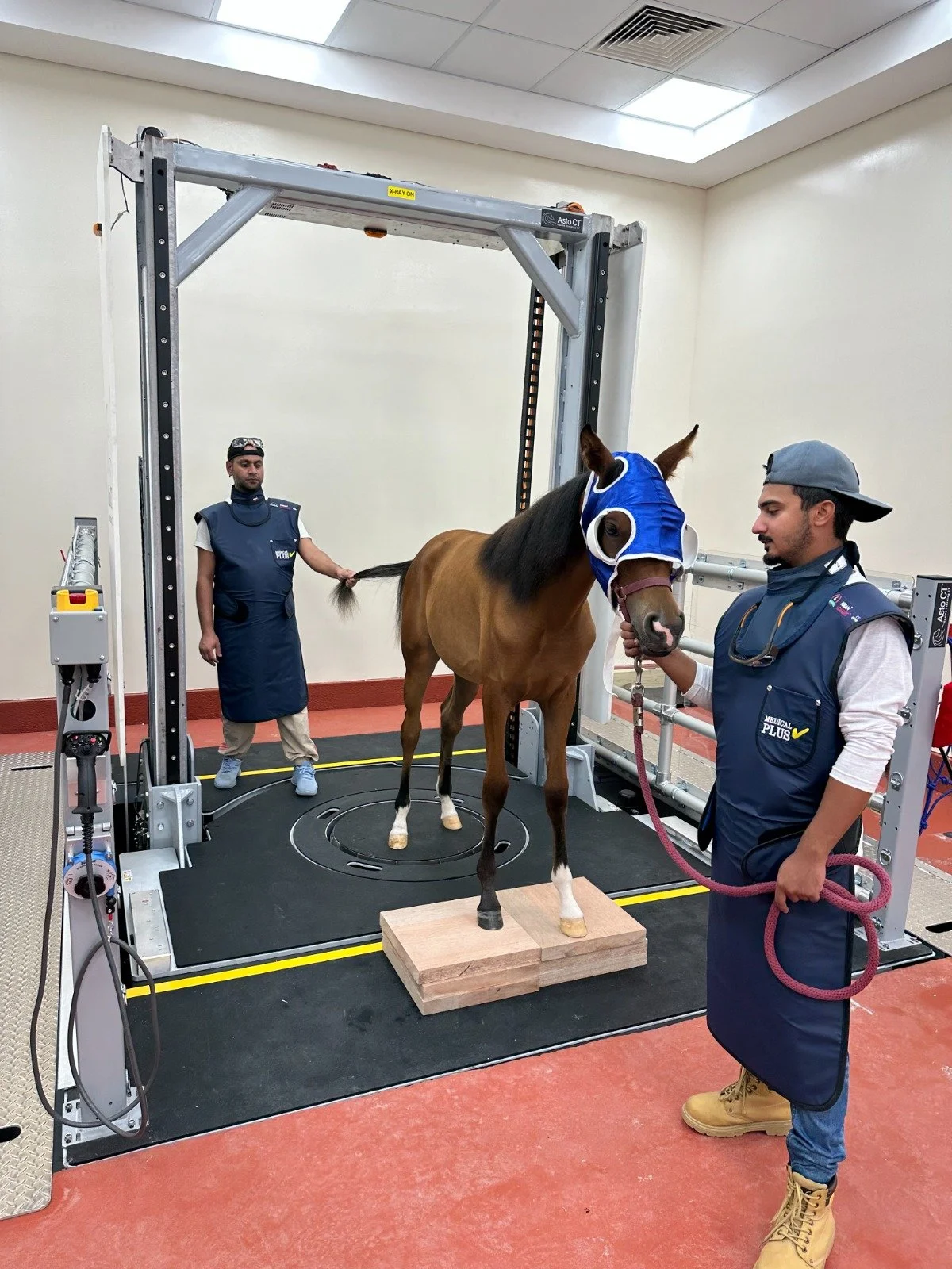
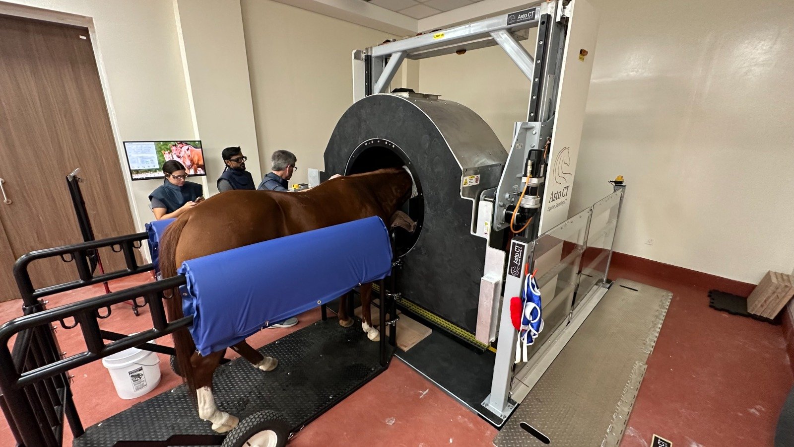
Revolutionizing Equine Healthcare: Sharjah Equine Hospital Introduces State-of-the-Art Equine Standing CT Scanner
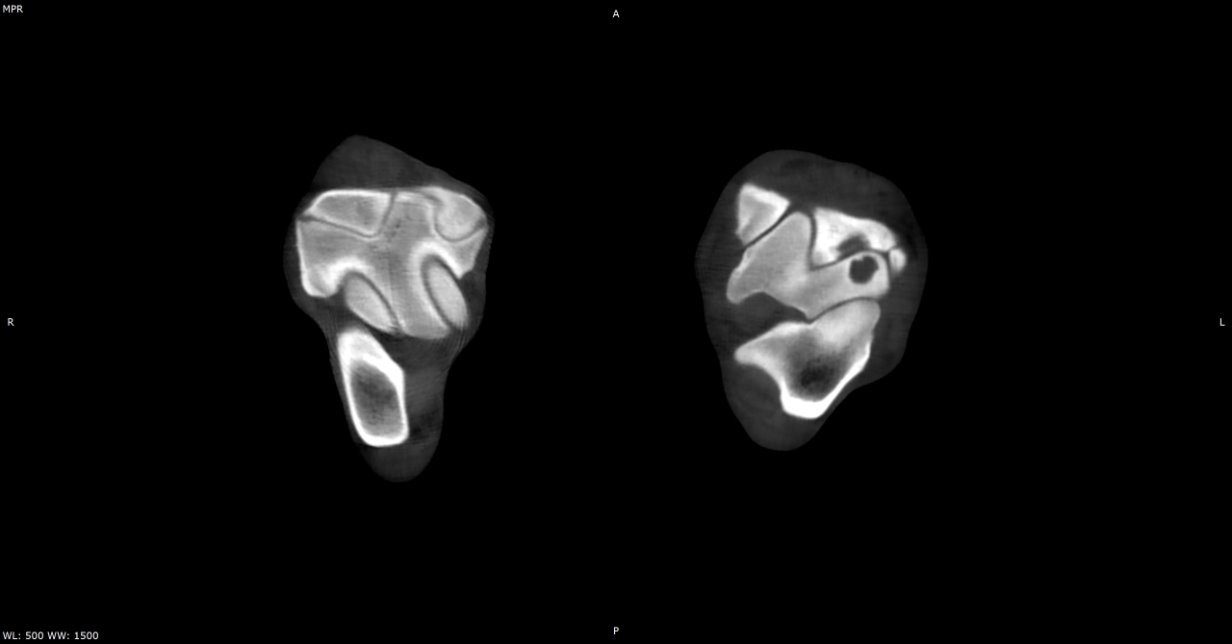
Case Study: 2-Year-Old Arabian Foal – Bone Cyst in the Lateral Trochlear Ridge of the Talus
Clinical History:
August 16, 2023: Diagnosed with osteochondritis dissecans (OCD) in the left hock. Underwent arthroscopic removal of a fragment.
September 19, 2023: Developed moderate to severe hock distension and 3/5 left hind limb lameness. Treated with intra-articular hyaluronic acid and triamcinolone, showing initial improvement.
Clinical Examination:
October 17, 2023: Worsened to 4/5 lameness and referred for further investigation.
Left hock showed moderate distension but was neither warm nor painful on palpation.
No significant reaction observed upon flexion.

Diagnostic Imaging:
Radiographs revealed a well-demarcated circular radiolucency on the proximal aspect of the lateral trochlear ridge of the talus, consistent with a subchondral bone cyst.
Case Study: 8-Year-Old Selle Français Mare – Luxation of the Right Temporohyoid Joint & Stylohyoid Bone Fracture
Clinical History:
The mare was referred after becoming stuck in a trailer with her neck bent to the right. Upon release, the owner observed superficial wounds, throat pain, and excessive drooling.

Clinical Examination:
Neurological signs:
Right ear and upper lip drooping
Absent menace response
Preserved pupillary reflex
Other symptoms:
Excessive salivation from the right side
No additional neurological deficits

Diagnostic Imaging:
Radiography: No abnormalities detected in the head or cervical vertebrae.
Endoscopy: Hematoma-like lesion extending along the stylohyoid bone within the right guttural pouch. The left guttural pouch appeared normal.
CT Diagnosis: Luxation of the right temporohyoid joint and a complete and displaced fracture of the stylohyoid bone.