
From Prototype to Clinical Implementation: Standing Computed Tomography in Equine Medicine
Introduction
Asto CT has performed over 10,000 standing computed tomography (CT) scans on horses worldwide. This milestone reflects more than a decade of development, engineering refinement, and clinical validation. The technology addresses a major limitation in equine imaging: traditional CT requires general anesthesia, which carries risks for injured or compromised patients.
Development and Early Prototypes
The system originated at the University of Wisconsin–Madison, where Dr. Peter Muir and Dr. Mark D. Markel sought to eliminate anesthesia for equine CT. They collaborated with Dr. Thomas “Rock” Mackie and medical physicist Dr. David Ergun to translate the concept into a clinical platform. Initial horse-tolerance testing used a full-scale wooden mock-up, confirming that standing scanning was feasible. The prototype was refined into a production-ready system through engineering improvements and extensive clinical feedback. Today, it is deployed in multiple countries and widely used in leading equine centers.
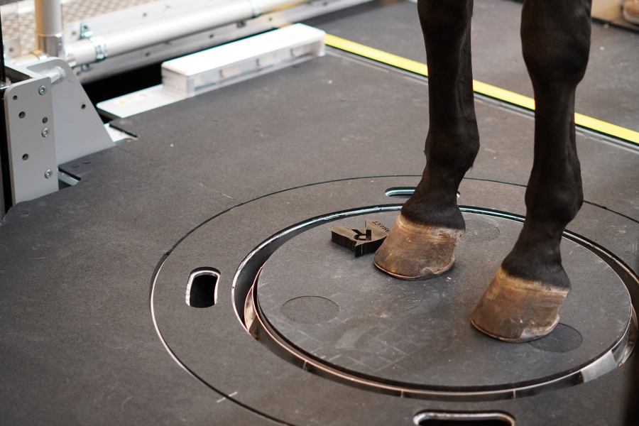
Figure 1: Image shows horse standing weight-bearing on CT platform.
Diagnostic Capabilities
The resulting Equina® system permits simultaneous imaging of both distal limbs in a weight-bearing stance (figure 1). This enables assessment of anatomical structures under natural load, improving detection of subtle pathology.
According to Clinical Professor and Asto CT Clinical Advisor Dr. Sabrina Brounts, the system’s high-resolution scans and three-dimensional reconstructions provide more diagnostic detail than radiography (Brounts et al.). The technology has proven effective for identifying fractures, subchondral bone lesions, and joint abnormalities. The ability to obtain accurate images without anesthesia reduces patient risk and shortens procedure time.
Applications in Injury Prevention
Standing CT is now part of established injury-prevention protocols in competitive equine sports. Since 2020, the University of Melbourne has used the Equina system for pre-race screening of horses entered in the Melbourne Cup. Before the system’s adoption, serious race-day injuries had occurred; following its integration, no major incidents have been recorded at the event (figure 2).

Figure 2: CT images of a carpus.
Such results underscore the system’s role in identifying early-stage pathology and informing evidence-based decisions on training, rest, or withdrawal from competition.
Melbourne Cup Safety Leadership Continues: Five Years of Standing CT Screening, Zero Fatalities
Infographic: Can We Predict Fetlock Stress Fractures Before They Happen?
Educational and Research Use
Standing CT is integrated into the curricula of multiple veterinary teaching hospitals for instruction in diagnostic imaging, lameness evaluation, and surgical planning. Its ability to produce high-quality images without anesthesia enables frequent use in both teaching and routine clinical case management. In research, eliminating anesthesia removes a major confounding factor, enhancing the validity of longitudinal and repeated-measure studies.
Eco-Friendly and Cost-Efficient Imaging
The Equina® Standing CT combines high-quality diagnostic performance with low energy use. Operating at 8 mA on a standard single-phase 208–240 VAC outlet, each 30-second scan consumes only 1.28 kW. For a clinic performing 150 scans annually, this amounts to 1.6 kWh, equivalent to the electricity needed to run a 9.5-watt LED light bulb for one week (source). Annual energy use could be offset entirely by a single 350-watt solar panel receiving five hours of sunlight2. The system’s compact footprint, under 18’ x 23’, and plug-and-play setup eliminate costly facility modifications typically required for conventional human CT scanners, providing clinics with sustainable, cost-effective advanced imaging.
Eco-Friendly CT Imaging: How the Equina® CT Saves Energy, Money, and the Planet

Figure 3: 11-month-old Holstein Bull receiving front limb scan with Equina® CT.
Adaptation to Other Species
Although designed for horses, the Equina platform has been adapted for other large and small animals (figure 3). Clinical cases include imaging of marine mammals, cattle, camelids, and small ruminants. For example, standing CT of an 11-month-old Holstein bull was successfully completed without anesthesia, demonstrating the system’s broader applicability (Means et al.).
Standing computed tomography is feasible and has clinical utility in ruminants
Sustained Collaboration
Long-term collaboration among veterinary clinicians, engineers, and industry partners has driven the advancement of standing CT. Feedback from real-world clinical use has guided hardware and software improvements, ensuring the system meets the operational demands of high-volume veterinary practices. Current development focuses on expanding anatomical coverage, improving image resolution, and integrating complementary modalities such as positron emission tomography (PET).
Conclusion
From a wooden prototype to over 10,000 scans completed, Equina® has evolved into a well-established clinical tool. It offers a safer alternative to recumbent CT for equine patients and has applications in diagnosis, injury prevention, education, and research. Its adaptability to other species further extends its relevance across veterinary medicine.
To read the full history, technical details, and case studies on Equina® standing CT, click here.
References
1. Brounts SH, Lund JR, Whitton RC, Ergun DL, Muir P. Use of a novel helical fan beam imaging system for computed tomography of the distal limb in sedated standing horses: 167 cases (2019–2020). J Am Vet Med. 2022;260(11):1351-1360. doi.org/10.2460/javma.21.10.0439
2. Means, K. L., Loeber, S., & Brounts, S. H. (2026). Standing computed tomography is feasible and has clinical utility in ruminants. American Journal of Veterinary Research, 87(1), Article ajvr.25.07.0254, ajvr.25.07.0254. Retrieved Mar 13, 2026, from https://doi.org/10.2460/ajvr.25.07.0254