
Equine Standing CT: Clinical Applications in Diagnosing Common Head and Cervical Pathology
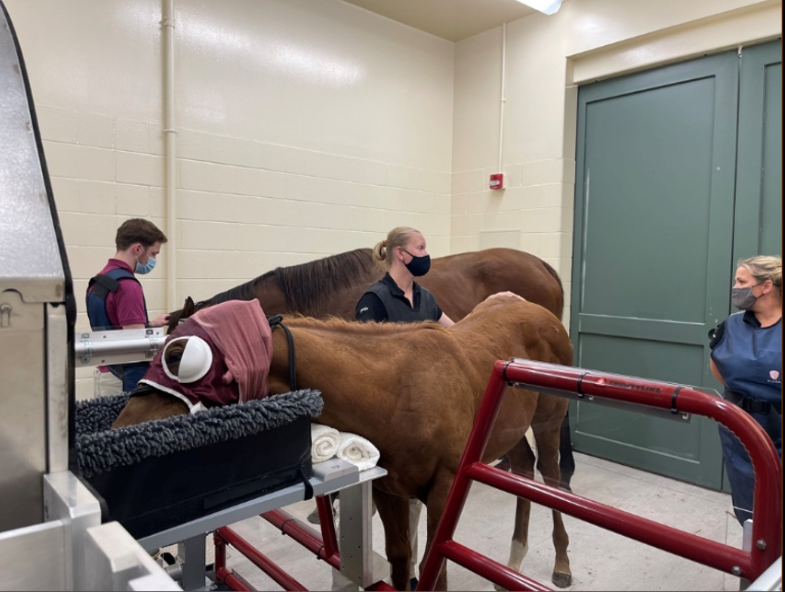
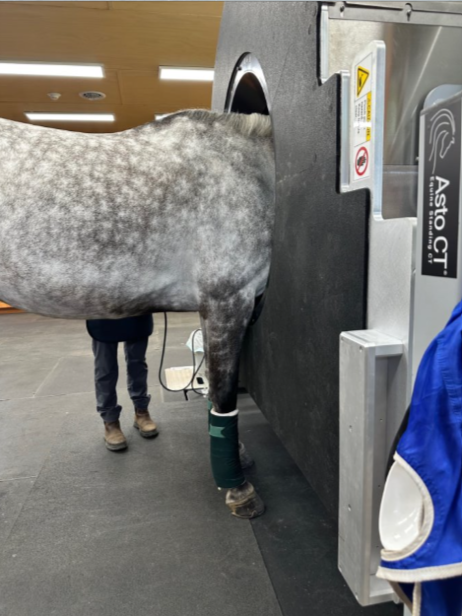
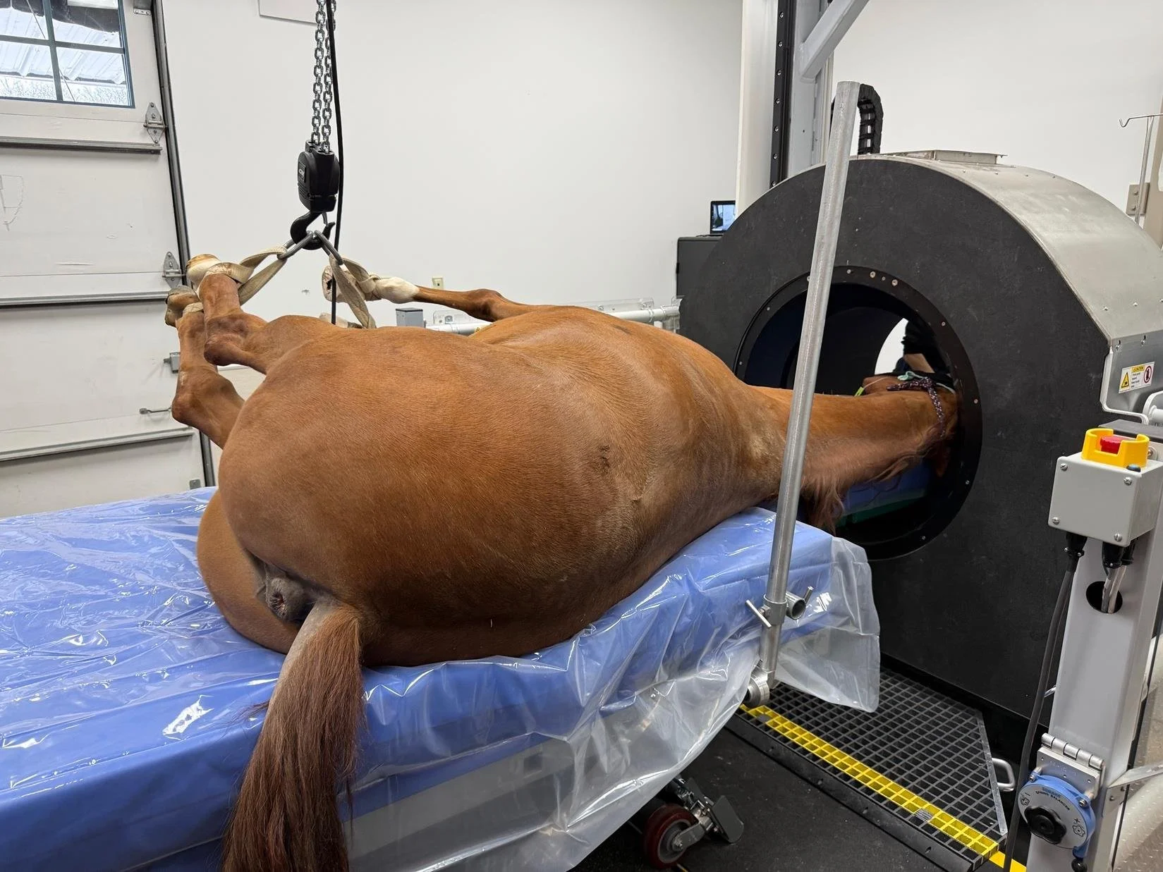
Advances in standing computed tomography (CT) have significantly expanded diagnostic capability in equine practice. The Equina® standing CT system provides high resolution, multiplanar, three-dimensional visualization of osseous and soft tissue structures of the head and cervical region under sedation. This reduces anesthetic risk and improves diagnostic accuracy across a wide range of pathologies. Recent developments, including a rear mounted headboard that allows consistent access to C6 and C7 and expanded options for imaging under general anesthesia, have further broadened clinical utility. The following review summarizes six categories of equine head and neck diseases in which standing CT using the Equine system has made a difference in diagnostic evaluation.
Skull & Mandibular Fractures
Case Example: 15‑Year‑Old Welsh Mare, Courtesy of Cotts Equine Hospital
History:
Multiple traumatic kicks to the head with concern over damage to left mandible and TMJ. The patient was dull with abnormal left optical reflexes and a droopy left ear/muzzle consistent with nerve damage, she was hypersalivating with difficulty swallowing and had an increased respiratory effort and inspiratory stridor. Therefore, a head CT was performed.
CT Findings:
Severely comminuted acute (mildly) displaced closed and non-articular fracture of the ramus/neck of the left mandible. There was no radiographic evidence of luxation nor joint incongruency of the left temporomandibular joint.
Small acute simple complete (mildly) displaced closed and non-articular fracture of the left paracondylar process.
Faint hypoattenuating line though the left side of the cranium (parietal bone/zygomatic arch), DDx: vascular channel considered most likely, less likely fissure.
Severe soft tissue swelling of the left side of the head (axial and abaxial), DDx: oedema, cellulitis, hemorrhage.
Moderate to severe narrowing of the pharynx.
Possible hematoma formation in the left guttural pouch, DDx: concurrent collapse due to abaxial soft tissue swelling.
Outcome
CT enabled accurate localization and classification of fractures and soft tissue compromise, guiding treatment planning and airway monitoring.


Sinus Disease / Dental Pathology
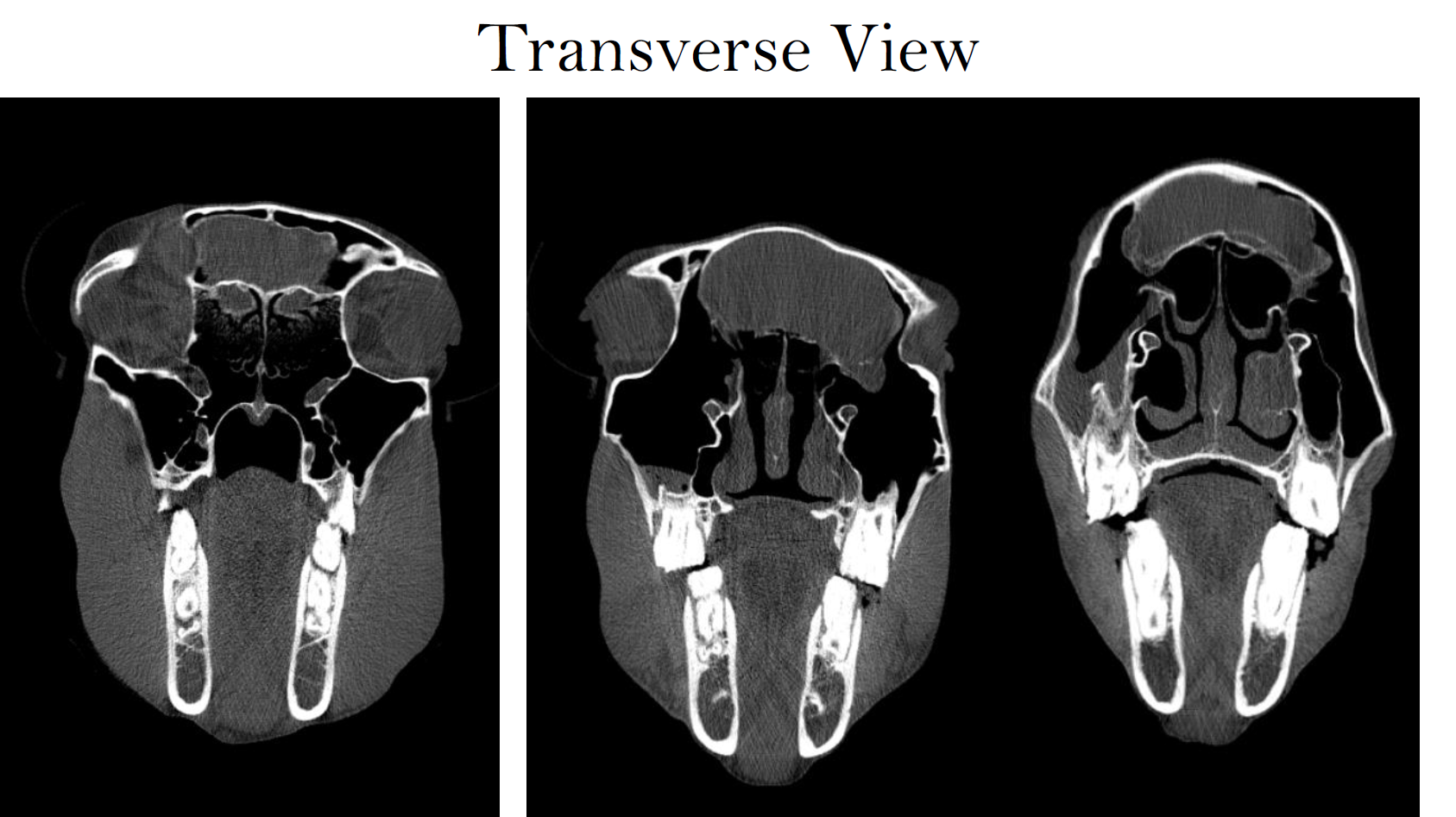
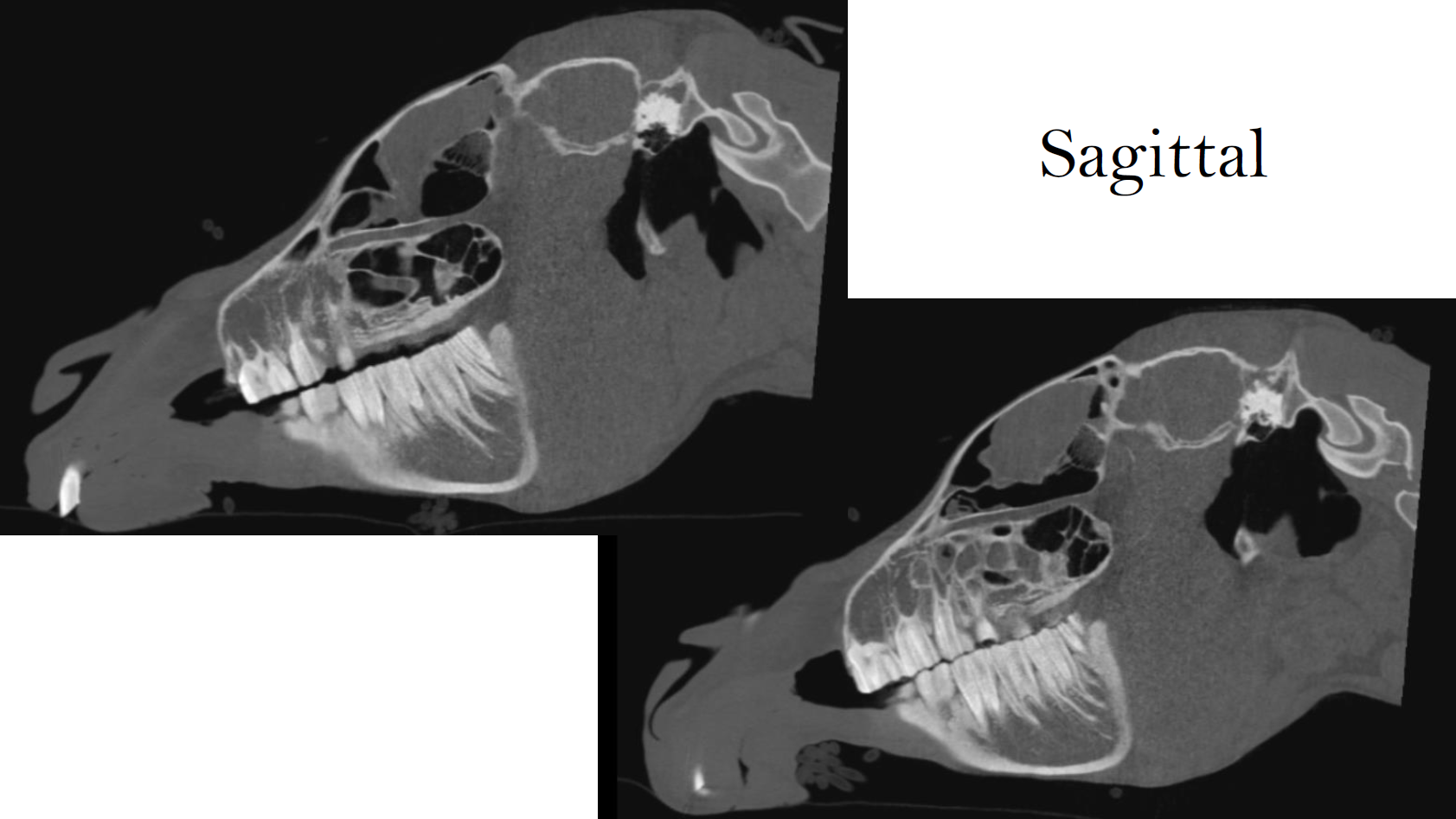
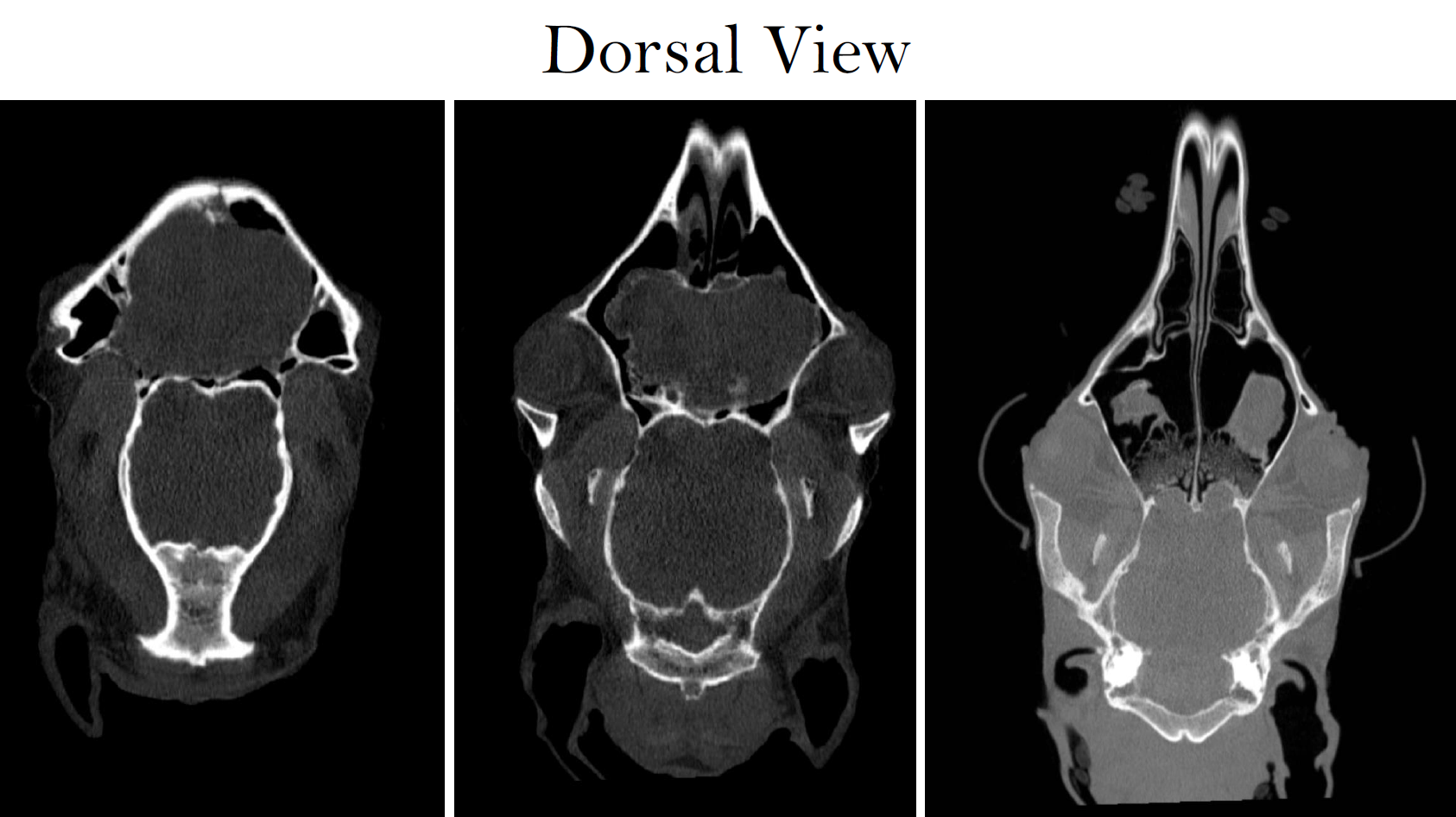
Case Example: 16‑Year‑Old Warmblood Mare, Courtesy of UW Large Animal Hospital
History
The mare presented with a firm, progressive swelling over the frontal sinus region and intermittent mild purulent discharge from the right nostril. Airflow remained normal bilaterally, and no cranial nerve deficits were present.
Presentation
Physical examination revealed a convex, firm swelling over the frontal sinus with mild right-sided nasal discharge. Facial symmetry and neurologic function were normal. Standing CT was pursued to characterize the lesion.
CT Findings
Imaging identified a peripherally mineralized concho frontal midline mass, consistent with a sinus cyst.
Additional findings included bilateral, asymmetric rhinitis, paranasal sinusitis, hyperostosis of the right maxillary alveolar bone (teeth 109–111), and mild odontoclastic tooth resorption with hypercementosis of the incisors.
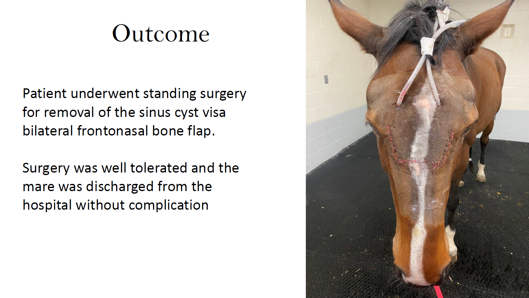
Outcome
A standing bilateral frontonasal bone flap procedure was performed, allowing complete cyst removal without complication. The mare recovered uneventfully and was discharged with an excellent prognosis.
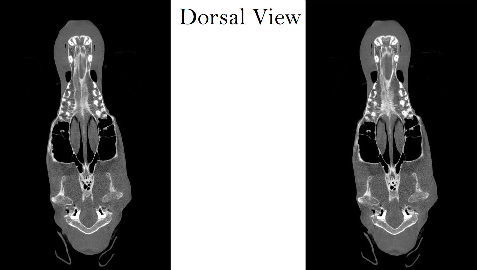
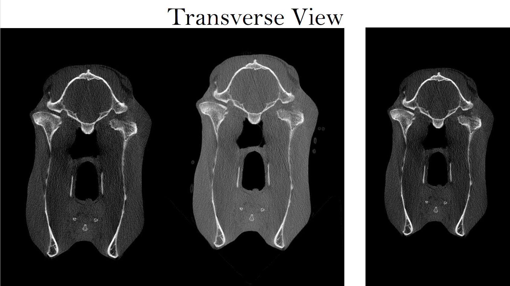
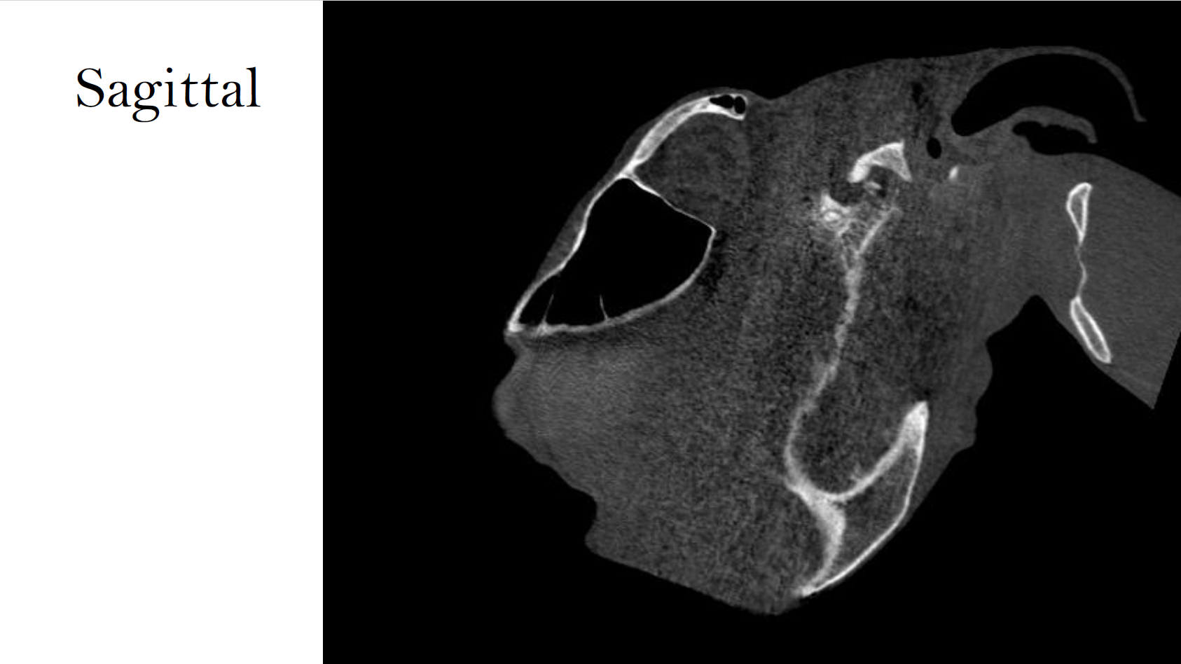
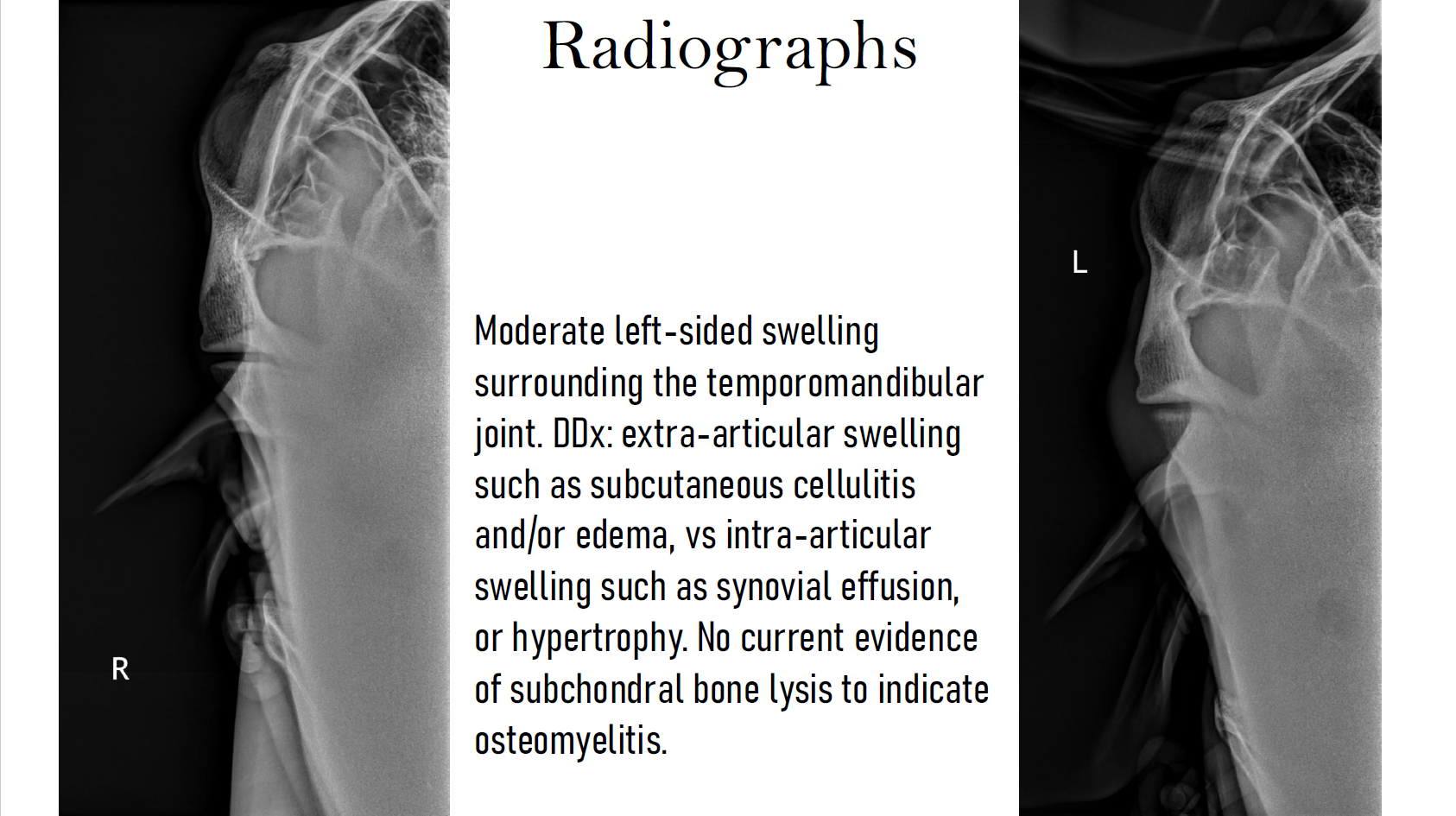
Temporomandibular Joint disease — Septic Arthritis
Case Example: 17‑Year‑Old Quarter Horse Gelding, Courtesy of UW Large Animal Hospital
History
The gelding was referred for worsening swelling, pain, and dysfunction of the left temporomandibular joint (TMJ). Clinical signs included lethargy, reduced feed intake, difficulty chewing hay, and marked sensitivity over the affected area.
Presentation
The gelding exhibited significant facial swelling, heat, and pain on palpation of left TMJ. He chewed hay slowly and occasionally quidded, though soft feed intake was normal. Initial diagnostics showed severe synovial inflammation and bacterial infection of the left TMJ. Evaluation of the contralateral TMJ was within normal limits.
CT Findings
CT demonstrated severe left septic TMJ arthritis, left condylar osteomyelitis
Chronic nondisplaced fracture with osseous sequestration, left otitis media, and Grade 3 bilateral temporohyoid osteoarthropathy.
Incidental focal sclerosis of the left occipital condyle and atlas were also present.
Outcome
Despite arthrotomy and lavage of the left TMJ, together with systemic and local antimicrobial therapy, the infection progressed and resulted in a grave prognosis. Left mandibular condylectomy was considered but deemed unlikely to restore comfort due extensive osteomyeltis extending into the stylohyoid bone of the left side. Humane euthanasia was ultimately elected.
Soft Tissue Pathology — Perilaryngeal Emphysema with Abscess
Case Example: 8‑Year‑Old Quarter Horse Gelding, Courtesy of UW Large Animal Hospital
History
The gelding had been hospitalized for seven days at another facility for colic treatment. Management included an indwelling nasogastric tube for oral fluid administration and several episodes of lunging. Diagnostic procedures performed were gastroscopy, upper airway endoscopy, proximal duodenal biopsy, an oral glucose absorption test, and a dental exam with float.
The colic resolved with medical therapy, but the horse subsequently developed pharyngeal swelling with crepitus, dyspnea, fever, and reduced appetite. A temporary tracheostomy was performed before referral.
Presentation
The horse arrived with diffuse subcutaneous emphysema of the pharyngeal and cervical regions, marked swelling, and reliance on a temporary tracheostomy. Endoscopy revealed a near complete airway obstruction from laryngeal edema, though no discrete perforation was visualized.d.
CT Findings
Standing CT revealed:
Extensive perilaryngeal and retropharyngeal emphysema extending ventrally along fascial planes
Intralesional feed material and/or abscessation within retropharyngeal tissues
Regional cellulitis, suggesting penetrating mucosal trauma or sealed perforation
Significant laryngeal compression with reduced arytenoid mobility
Tracheitis and tracheal wall edema, likely related to the tracheostomy tube
Tracking cervical emphysema approaching the vertebral canal
Incidental right maxillary sinusitis
Outcome
Intensive antimicrobial therapy and supportive care led to progressive resolution of emphysema and abscessation. Laryngeal function normalized, swelling resolved, and the gelding returned to normal eating and behavior. Outpatient airway re‑evaluation was planned.


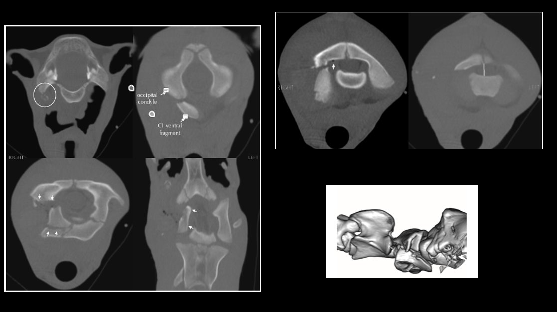
Cervical Vertebral Trauma
Case Example: 2‑Month‑Old Quarter Horse Colt, UMN Leatherdale Equine Center
History
The colt was discovered 36 hours after a suspected halter-related trauma with mild right head tilt, intermittent depression, and swelling over the left cranial neck and head.
Presentation
On referral, the colt was bright and neurologically appropriate. Examination revealed mild head tilt, palpable crepitus over suspected fracture area, normal gait at walk and trot, and no overt neurologic deficits. CT was performed to evaluate suspected cervical fracture.
CT Findings
CT identified fractures of the right jugular process and the right lamina of C1, with an open C1 fracture allowing a small amount of gas to enter the vertebral canal.
Fragment displacement caused moderate canal narrowing at C1–C2 and subluxation of the right atlanto‑occipital joints.
Soft tissue within the vertebral canal suggested hemorrhage or inflammatory debris, and mild spinal cord compression was suspected.
Outcome
The CT findings provided definitive localization and severity grading, guiding further decision-making regarding stabilization, prognosis, and monitoring.
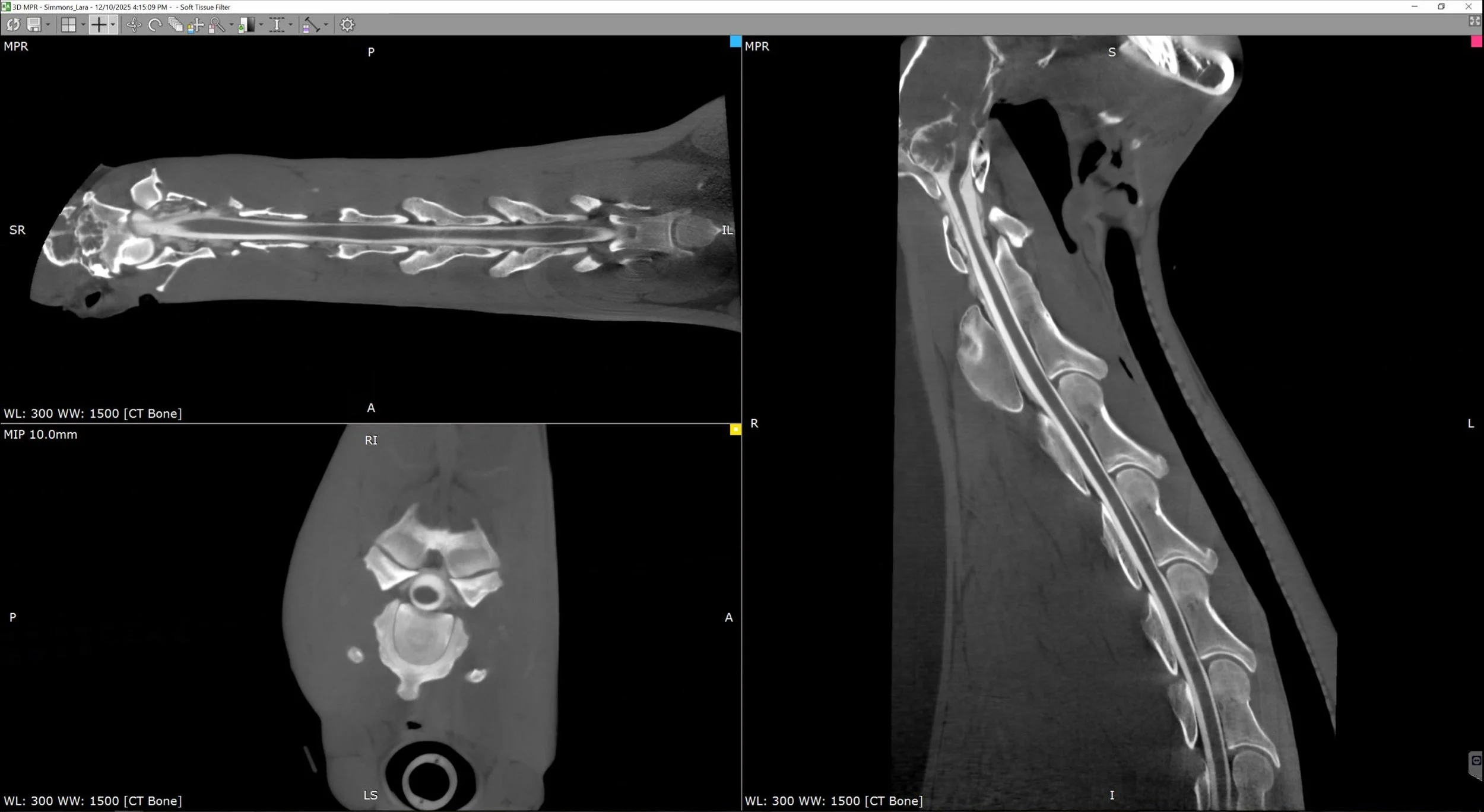
Cervical Vertebral Disc & Canal Abnormalities
Case Example: 5‑Year‑Old Thoroughbred Gelding, Courtesy of Cotts Equine Hospital
History
The gelding experienced an acute episode of severe pain followed by collapse and violent thrashing. He had been performing normally prior to the event. Examination revealed grade 3–4 hind‑limb ataxia, with the horse nearly falling when turning. Radiographs and ultrasound did not reveal a cause, prompting CT evaluation.
Presentation
Despite significant ataxia, standing CT successfully imaged the cervical vertebrae through the C5–C6 region.
CT Findings
Findings included:
Mild funnel-shaped narrowing of the vertebral canal at C4–C5
Lateromedial elongation and dorsoventral shortening of cranial C4 and C5
Flattened spinal cord appearance, with loss of surrounding epidural fat
Moderate dorsal lamina extension (C3–C5)
Mild endplate flaring (C3–C4)
Mild periarticular spur formation (C2–C6)
Mild buttress formation of the right cranial articular process of C6
Narrow intervertebral disc spaces (C2–C5)
Reduced intra‑vertebral sagittal ratios at C4 and C5 (<0.50), consistent with cervical canal stenosis
Outcome
The CT findings provided critical diagnostic information to guide neurologic prognosis and management, clarifying the structural abnormalities contributing to the horse’s severe ataxia.


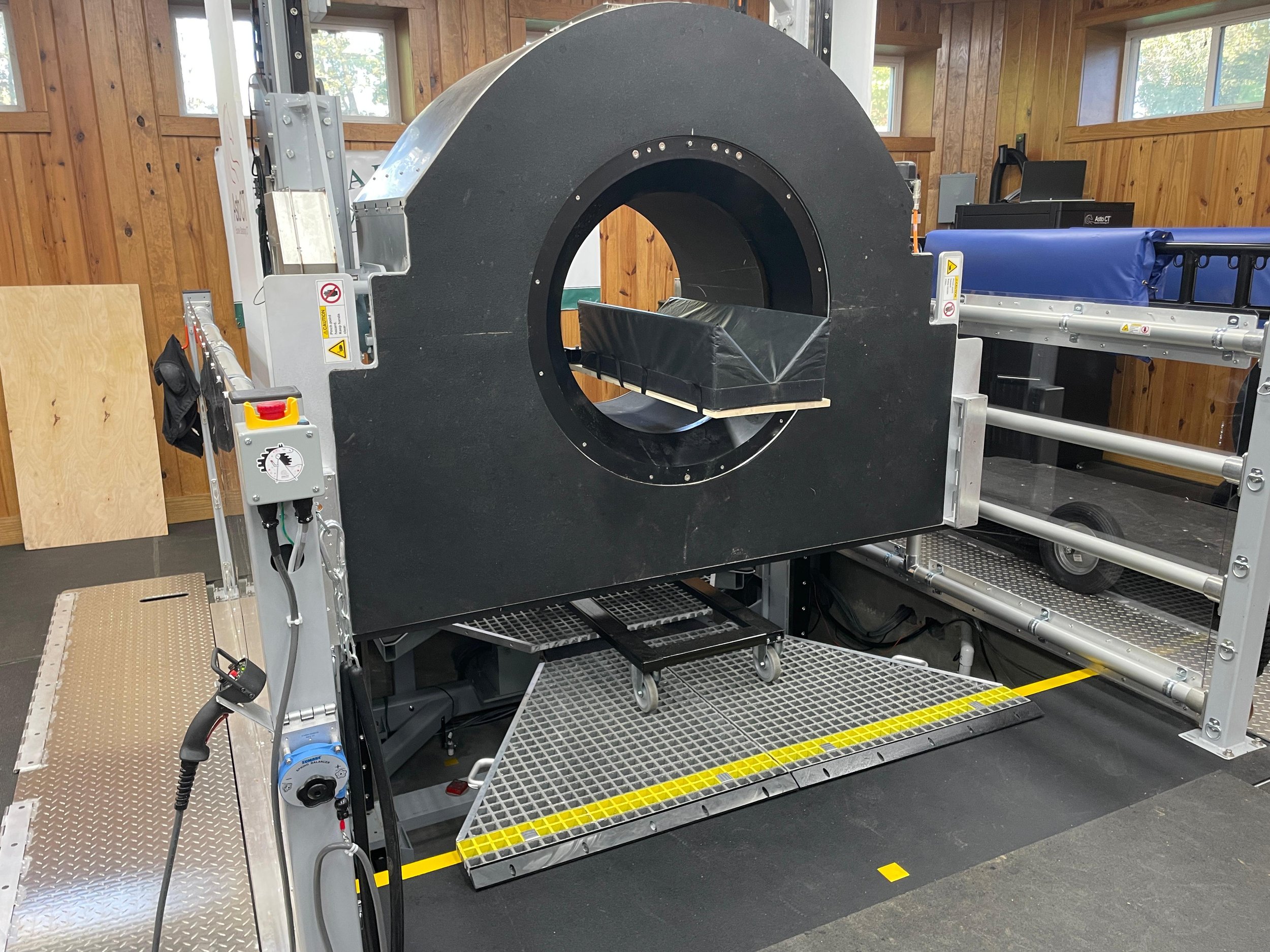
General Anesthesia Imaging Capabilities
Although designed for standing sedation, the Equina® platform can be used for recumbent imaging when necessary. Asto CT has developed an 80-centimeter surgical table extension that allows rapid and safe positioning of horses under general anesthesia. The extension supports loads up to 750 pounds and has been used clinically to complete cervical imaging from induction to recovery in less than 20 minutes. Imaging in lateral recumbency can provide visualization through C7 and T1 depending on conformation.


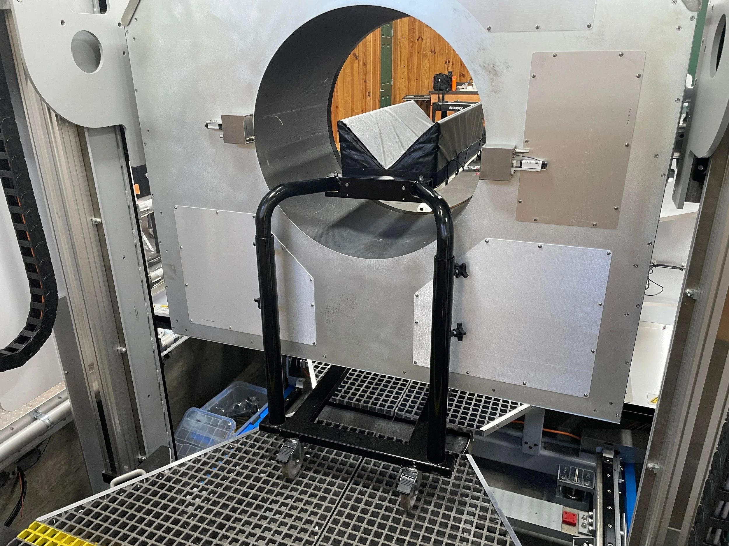
Access to Caudal Cervical Spine (C6 and C7)
With the rear mounted headboard and revised head support, clinicians can now consistently image the caudal cervical region including the cranial aspect of C7 in standing horses. This capability has been confirmed through studies performed at multiple clinical sites.

CT Cervical Myelography under general anestesia
The Equina® system supports CT myelography for evaluating spinal cord compression, extradural attenuation, and canal stenosis. The case of a 7-year-old Quarter Horse gelding demonstrates excellent contrast filling of the subarachnoid space with high-resolution three-dimensional reconstructions. These images provide clear definition of the cervical cord contour and canal margins.
CT myelography increases diagnostic sensitivity in neurologic cases where CT imaging alone may not demonstrate cord compression sufficiently.
Conclusion
Equine standing CT has reshaped the diagnostic approach to head and cervical pathology. Recent improvements to the Equina® system, including enhanced access to the caudal cervical spine, expanded general anesthesia imaging capabilities, and integration of CT myelography, continue to broaden the range of cases that can be evaluated effectively. These capabilities improve diagnostic accuracy and enable earlier, more targeted clinical intervention.
.