Scroll
Trending Research & Whitepapers
Featured
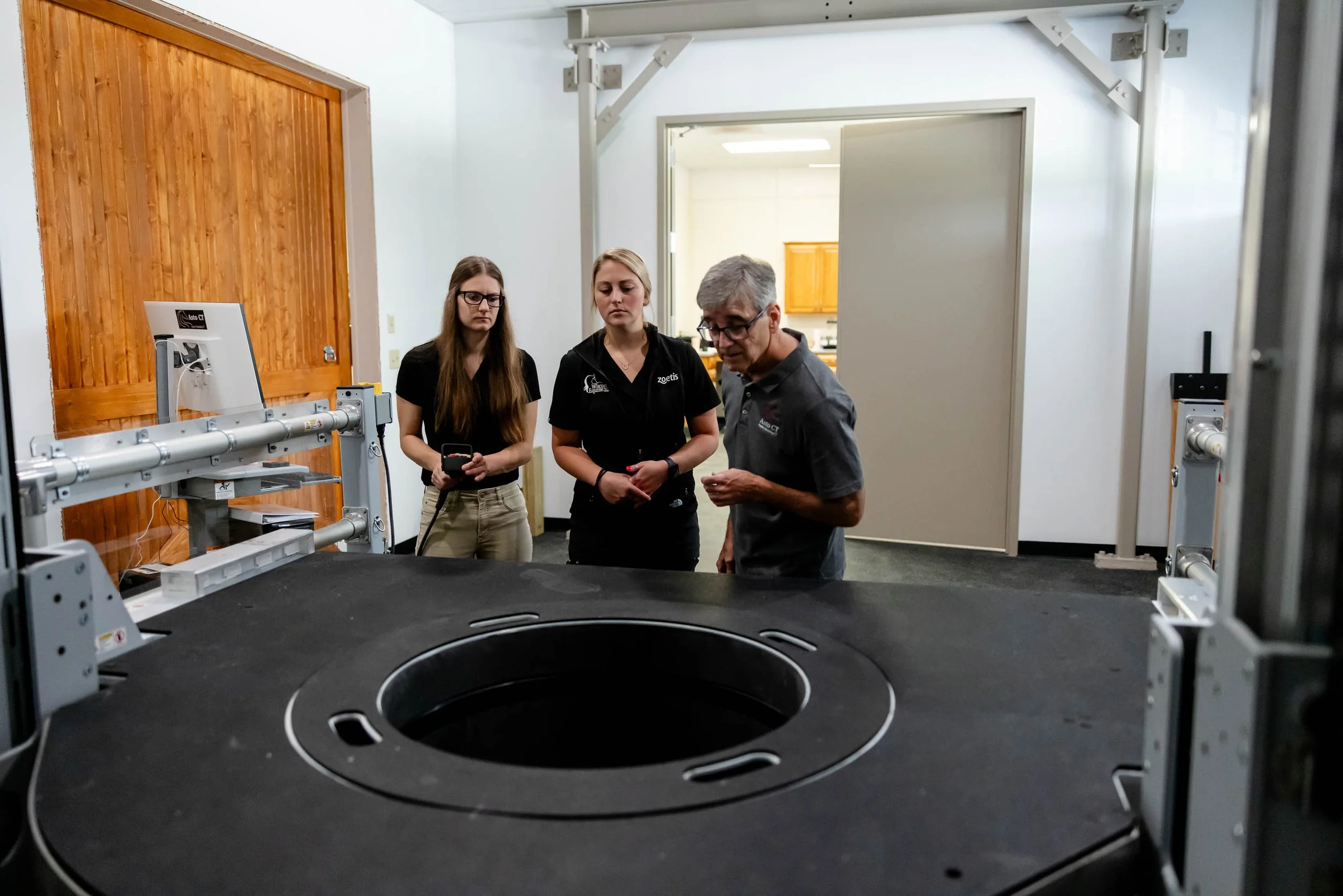
Our findings suggest that scanning large numbers of horses per year with the Asto CT Equina would not lead to occupational radiation exposure above the recommended safe threshold for handlers using lead shields and eyeglasses.
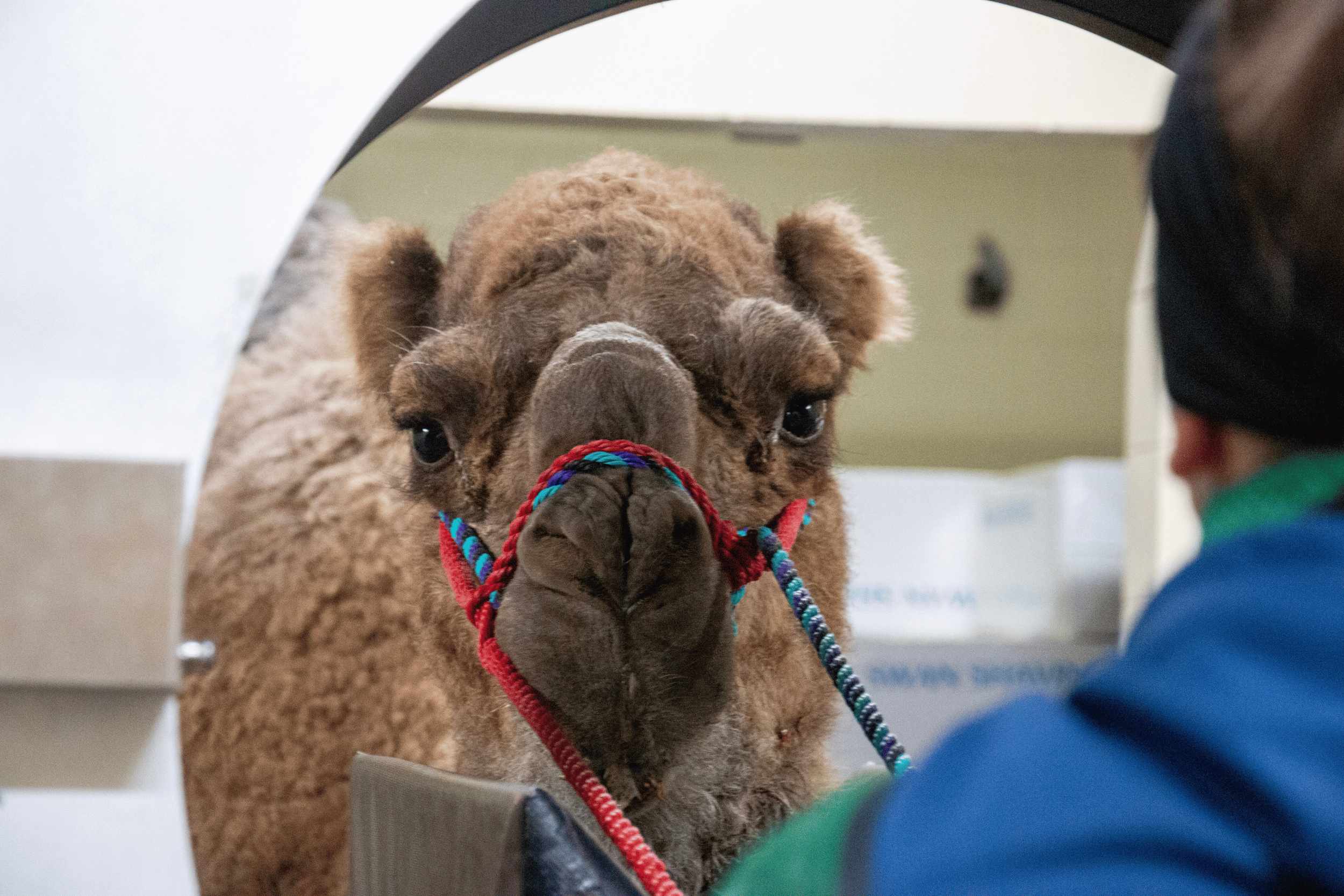
Sedated standing CT of the head and distal limbs is feasible in ruminants and may be an option in patients that are amenable to a standing imaging procedure.

CT imaging of the head of sedated standing horses has been evaluated in several studies, but studies involving CT imaging of the distal limbs have been more limited.

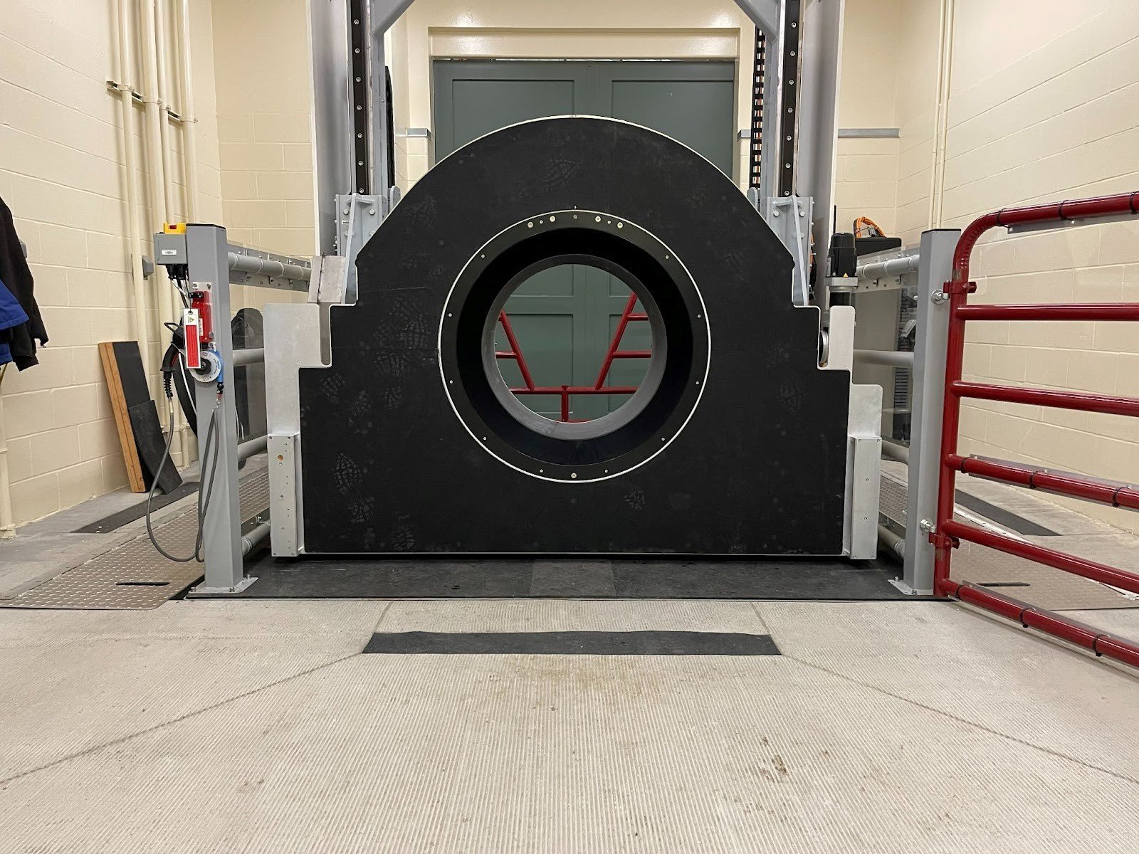
In the equine space, fan-beam systems offer several key advantages. Fan-beam systems offer faster scan times which make its images less susceptible to motion artifacts due to swaying or other subtle movements of the equine patient. Fan-beam images also have higher image quality, enabling them to be used to identify subtle pathological changes in the limbs of horses, and important advantage over both traditional radiography and cone-beam CT scanning.

The use of this advanced veterinary technology for early detection and management of musculoskeletal injuries can move us towards a new standard of care for Thoroughbred racehorses. In this paper, we provide a brief description of the key features of the Equina® system, and its use in pre-race screening.
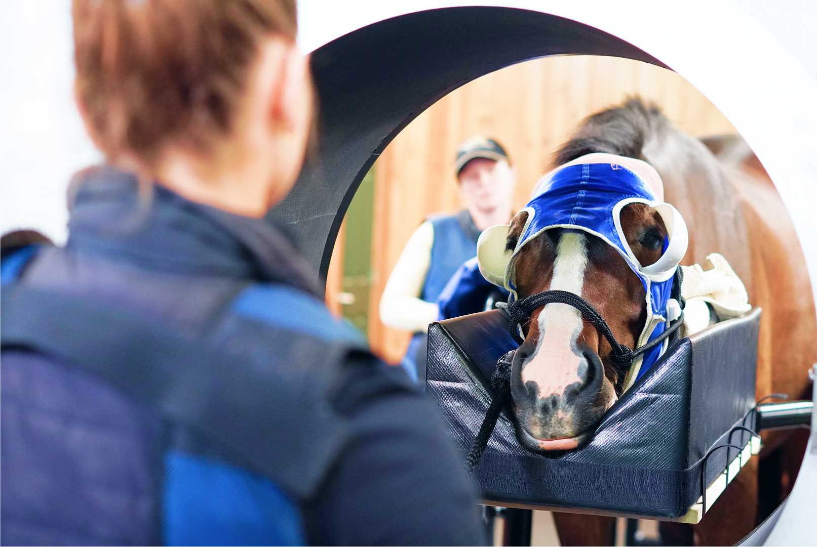
CT has revolutionized equine clinical practice over recent decades, with CT imaging of the equine head currently considered preferable to conventional radiography.
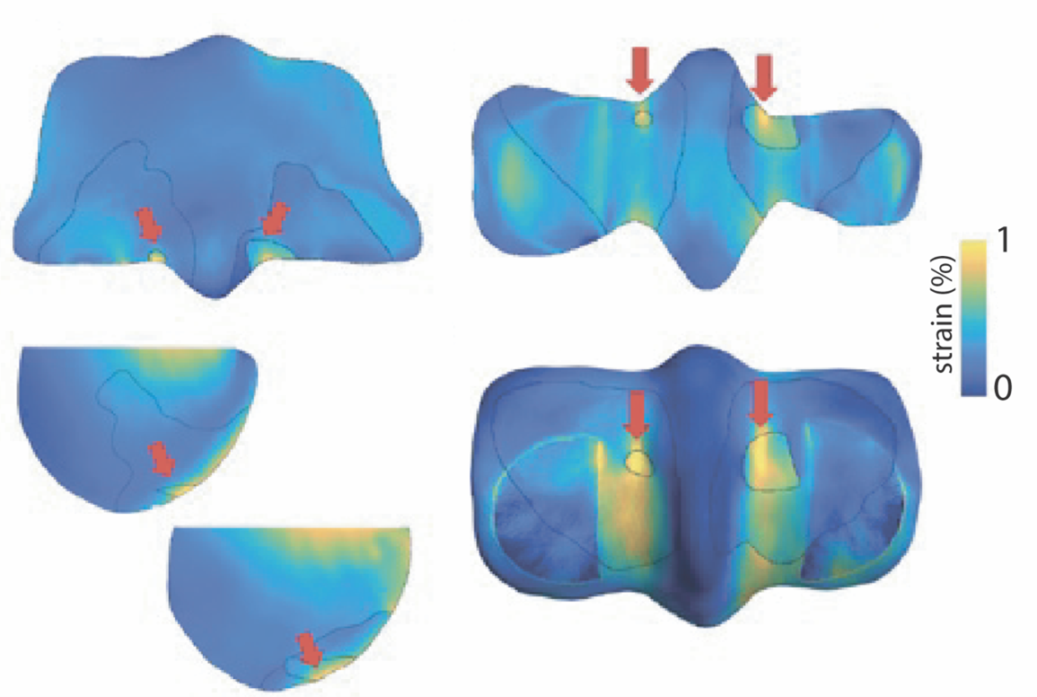
Condylar stress fracture of the third metacarpal bone (MC3) is a common catastrophic injury in Thoroughbred racehorses and is associated with parasagittal groove (PSG) subchondral osteolysis.
Discover Asto CT Articles
Featured
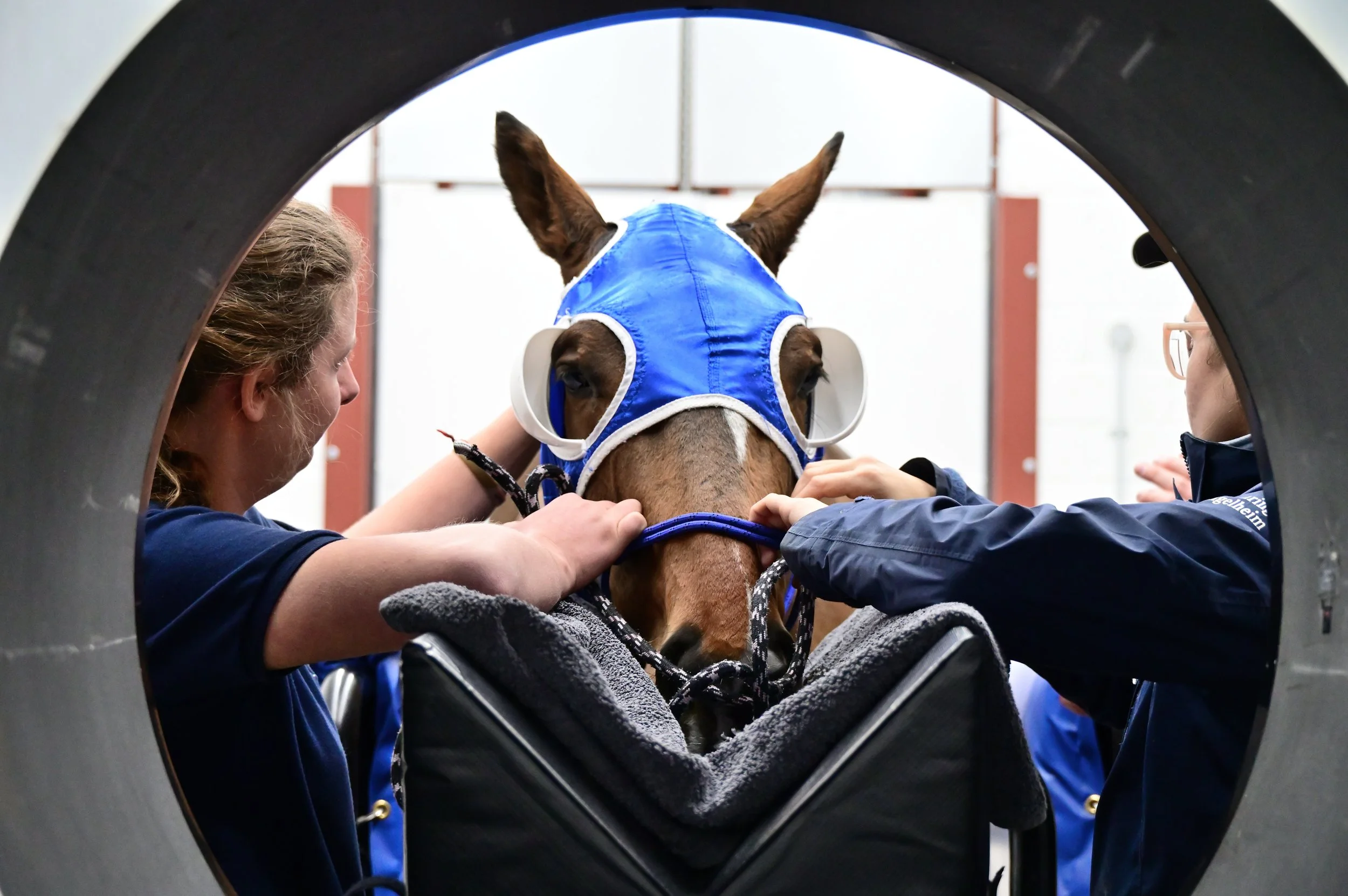
Why purpose-built equine imaging matters for the horse, the clinical team, and the practice. Read the article to learn what to consider before investing in a standing CT scanner.
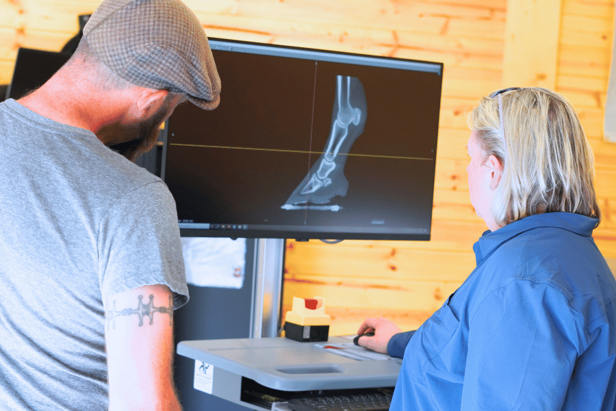
It makes sense that any equine practice with an advanced lameness program would have Equine Standing CT installed. For us at Virginia Equine Imaging, it’s a critical tool for diagnosing lameness and developing treatment plans…
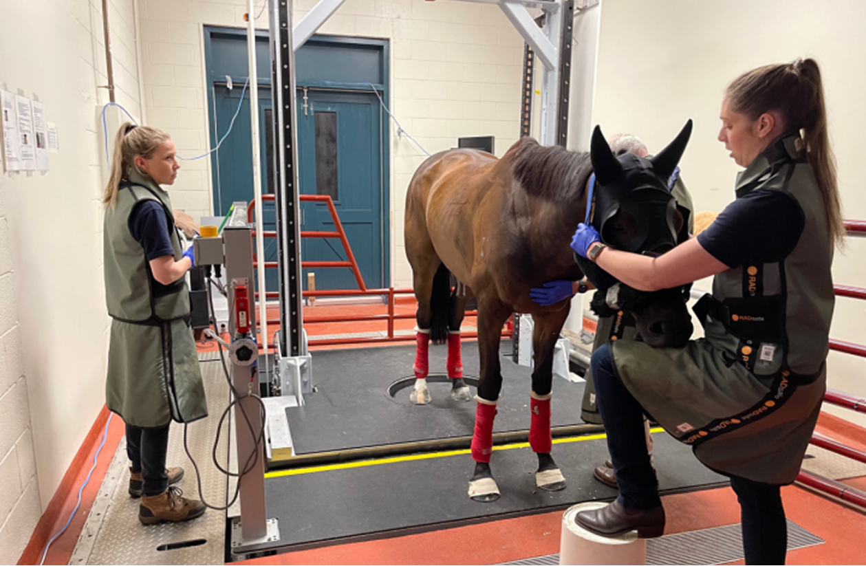
What began as a response to an unacceptable injury history has evolved into one of the most comprehensive pre-race veterinary screening programs in global racing, combining advanced imaging, longitudinal clinical assessment, and independent expert review.
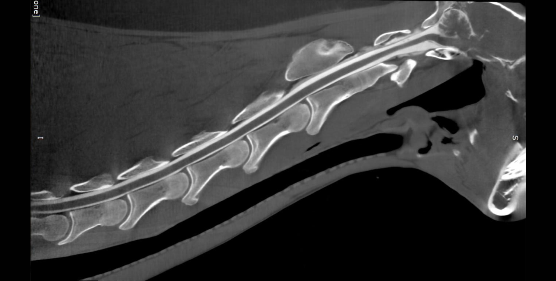
Standing CT continues to reshape how we evaluate challenging head and neck conditions in horses. This case‑based review highlights where CT meaningfully changes clinical decision‑making—from acute trauma to neurologic disease.
Can we predict fetlock stress fractures before they happen? New research in Equine Veterinary Journal shows how standing CT combined with virtual mechanical testing can assess fracture risk in elite Thoroughbreds by measuring strain in high-risk regions of the distal MC3.

The resulting Equina® system permits simultaneous imaging of both distal limbs in a weight-bearing stance. This enables assessment of anatomical structures under natural load, improving detection of subtle pathology.
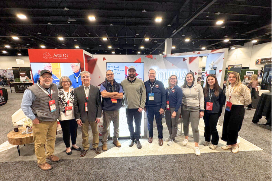
We are proud to announce the winners of the 2025 Asto CT Best CT Cases of the Year, recognizing exceptional clinical cases that highlight the diagnostic power, versatility, and clinical impact of standing CT imaging across equine and veterinary medicine.

“The Equine Standing CT at Mizzou is transforming how my colleagues and I diagnose and treat lameness,” says Dr. McCraken. “We’re identifying lesions in new locations and gaining a deeper understanding of how these findings correlate with clinical signs.”
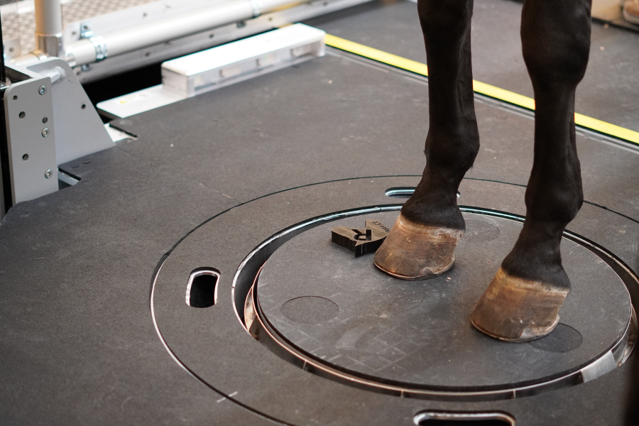
Bones, joints, and soft tissues align differently when a horse is non-weight-bearing. Weight-bearing CT captures the structures as they function during movement and while bearing load, leading to more accurate diagnoses.

Pioneer Equine Hospital, a premier, full-service equine veterinary practice proudly announces the integration of the cutting-edge Asto CT Equina® Standing CT Scanner into its diagnostic services. This state-of-the-art imaging technology marks a significant advancement in the hospital’s commitment to providing exceptional equine care.
Asto CT Webinars
Featured
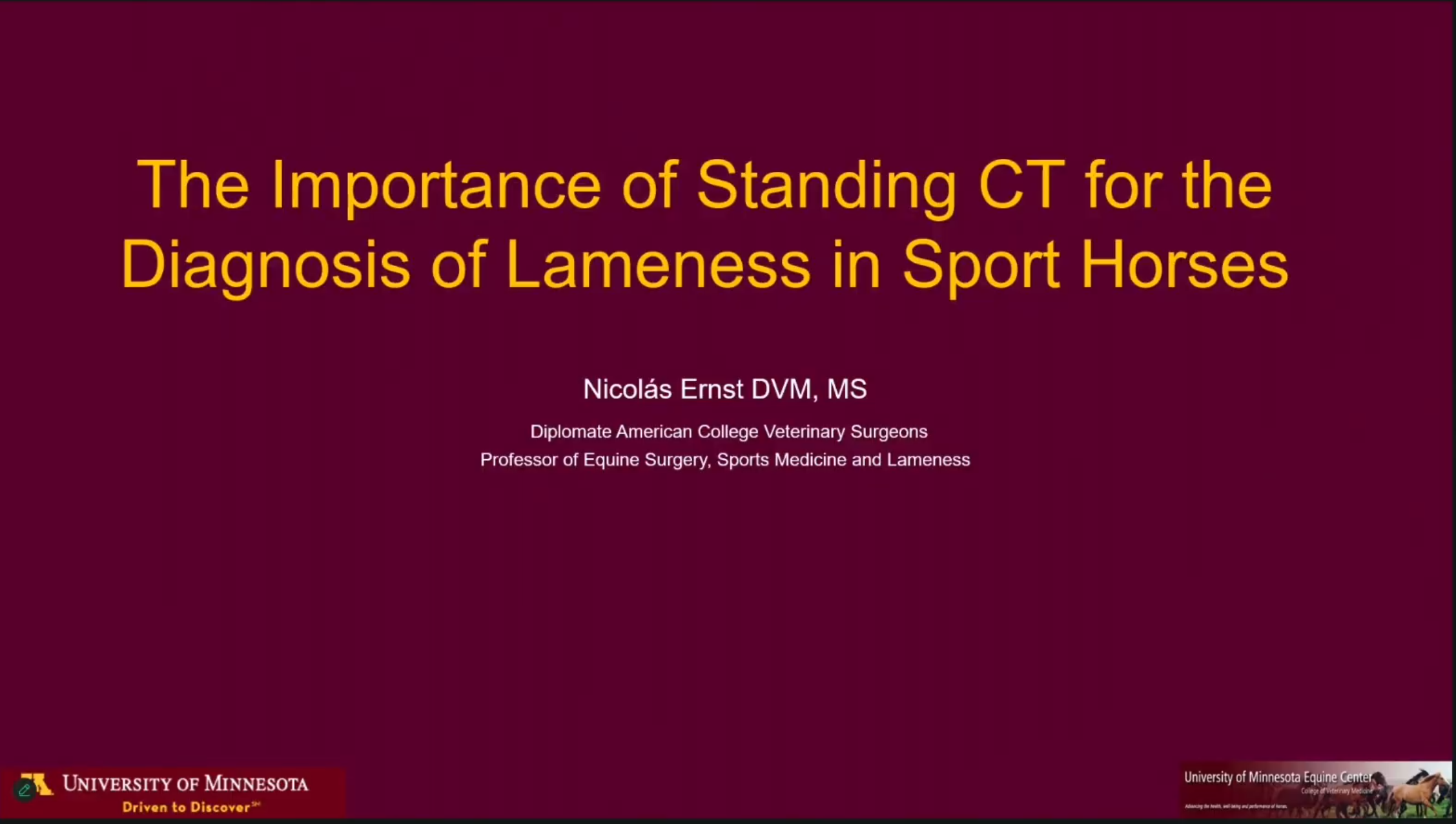
Asto CT Download Center
In this Planning Guide, learn:
How to select the ideal partner for your equine CT journey
Key milestones in installing an equine standing CT and ensuring timely delivery
The importance of choosing a CT partner committed to ongoing innovation and future upgrades
Or download our brochures below.

Visual Proof
Featured Case Studies
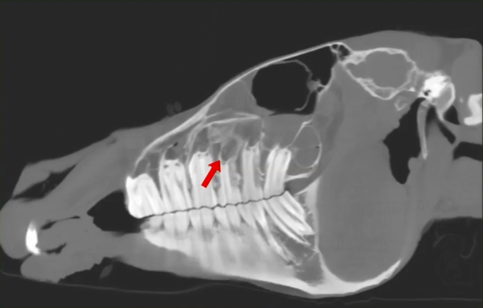
A 10-year-old TB mare was referred for evaluation of persistent right‑sided purulent nasal discharge that failed to respond to antibiotics. Despite significant nasal drainage, upper airway endoscopy was unremarkable, leaving the underlying cause unclear.
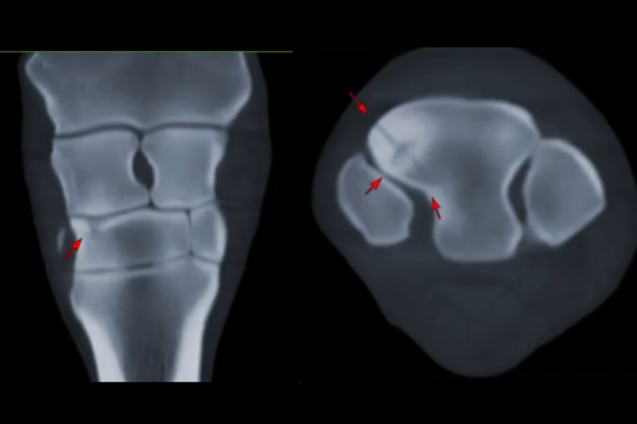
Traditional radiographs (including a skyline view) showed subtle changes but made the fracture difficult to visualize clearly. CT imaging provided a dramatic improvement in diagnostic clarity. The scan revealed a distinct fracture line, surrounding sclerosis, and areas of demineralization features that were not easily discernible on radiographs.
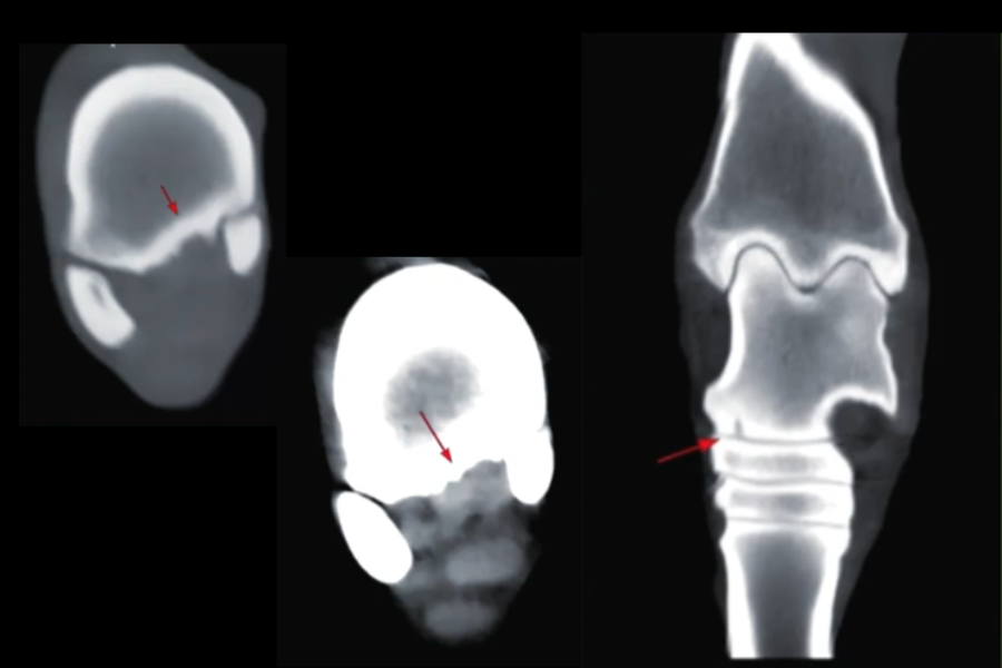
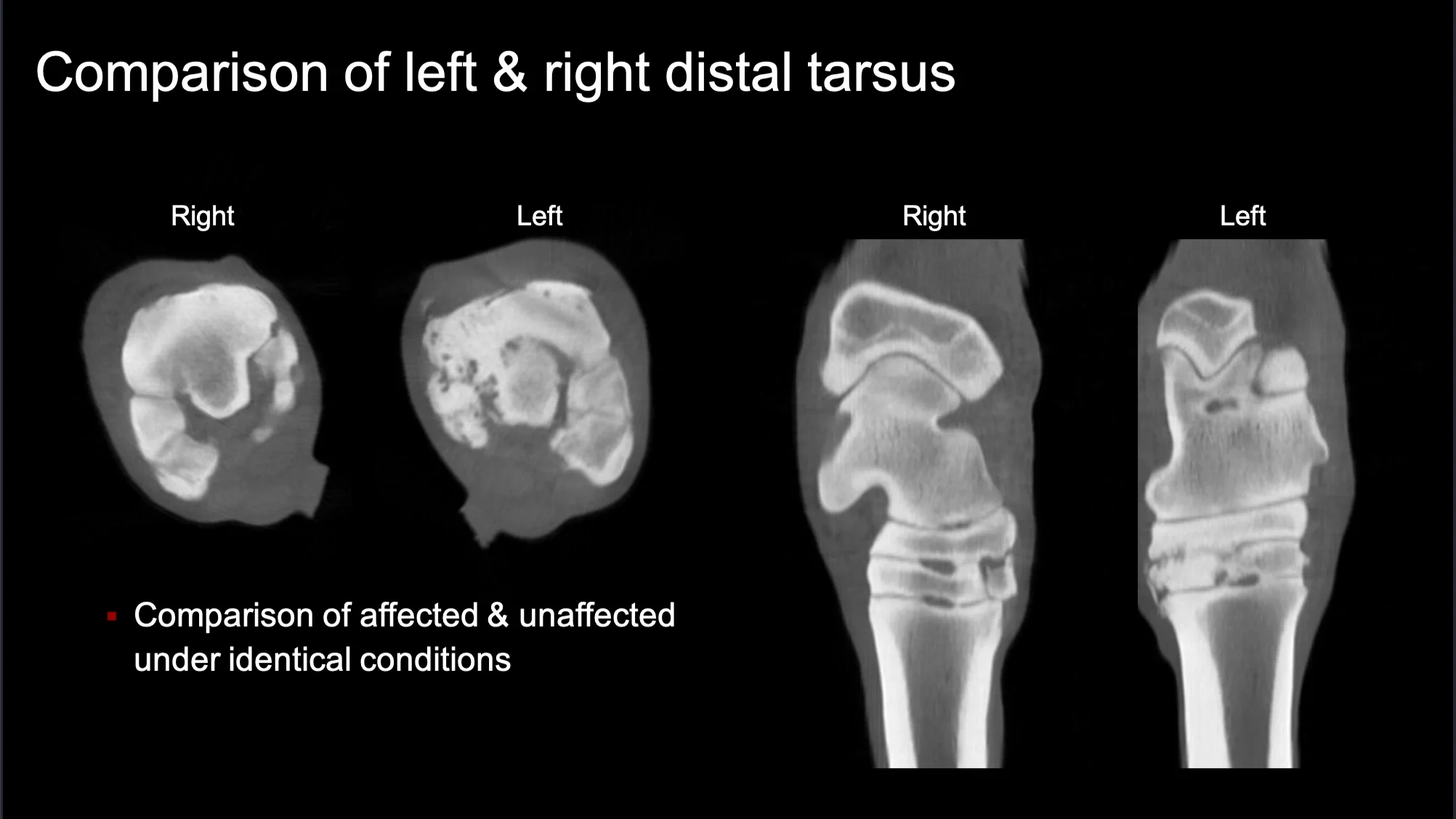
Standing CT revealed what radiographs did not: a distal medial talus subchondral defect bordered by sclerosis within the talocalcaneal region.
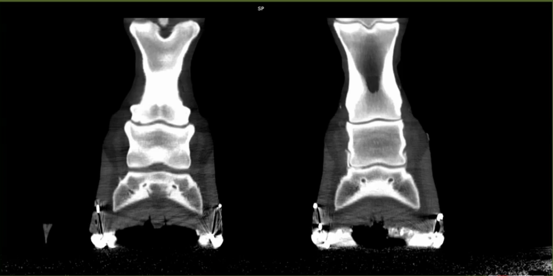
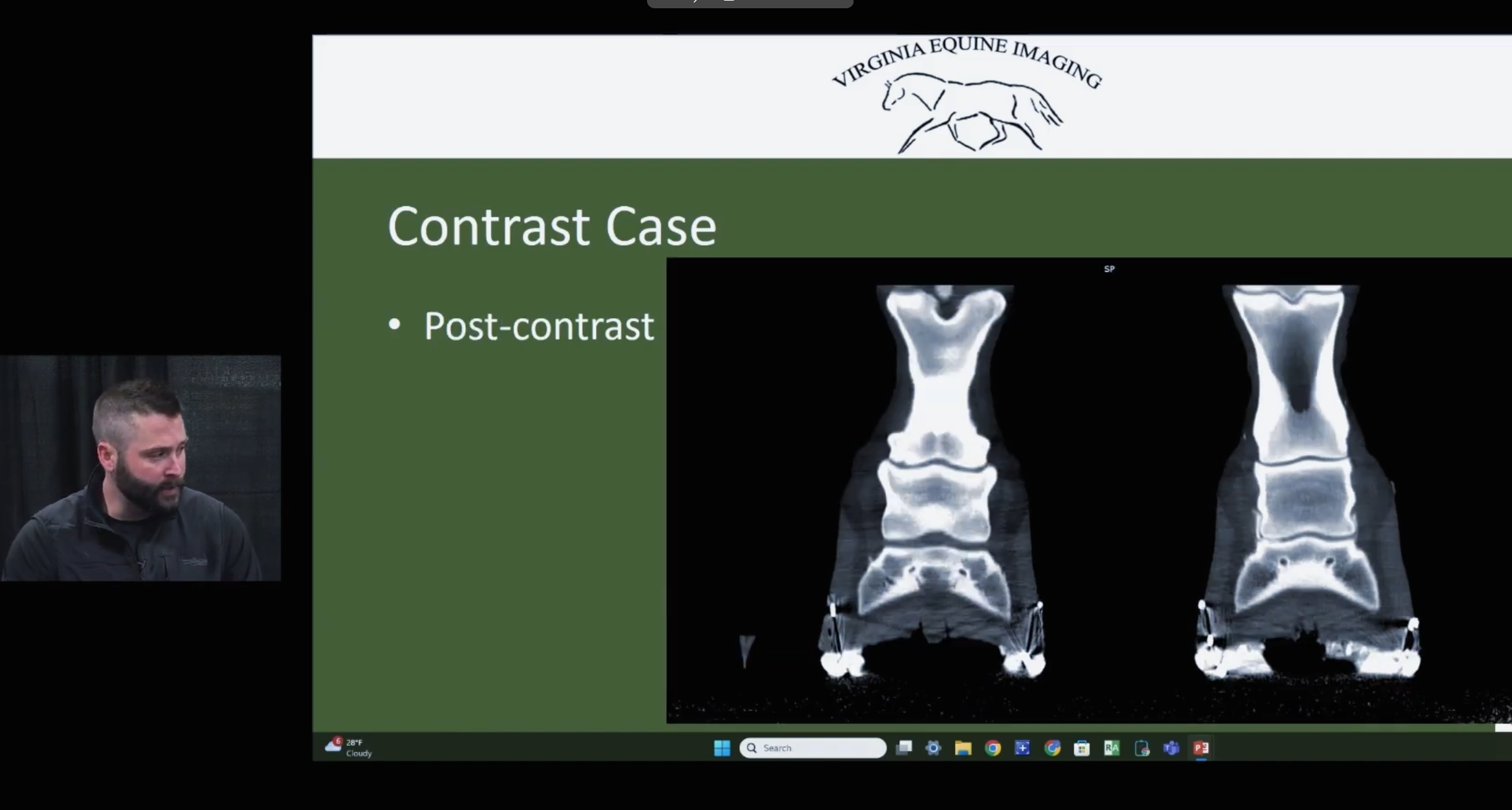
This case highlights the diagnostic power of CT with intra‑articular contrast for accurately assessing distal limb pathology, especially when MRI findings are equivocal or incomplete. CT provided a definitive understanding of the extent of cartilage loss and joint disease, ultimately guiding appropriate case management.
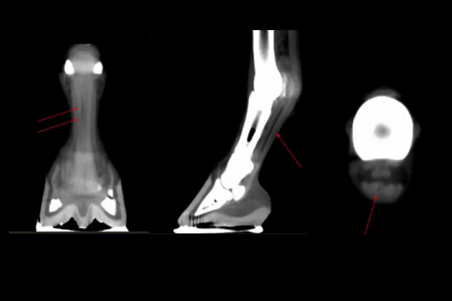
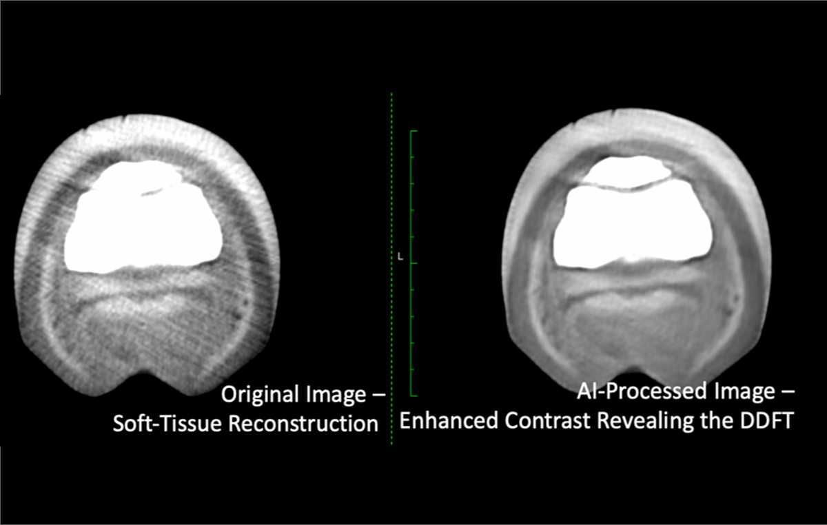
CT imaging revealed a moderately progressive DDFT tear now extending from mid‑P1 to the distal navicular bone, including a new full‑thickness parasagittal suprasesamoidean tear.
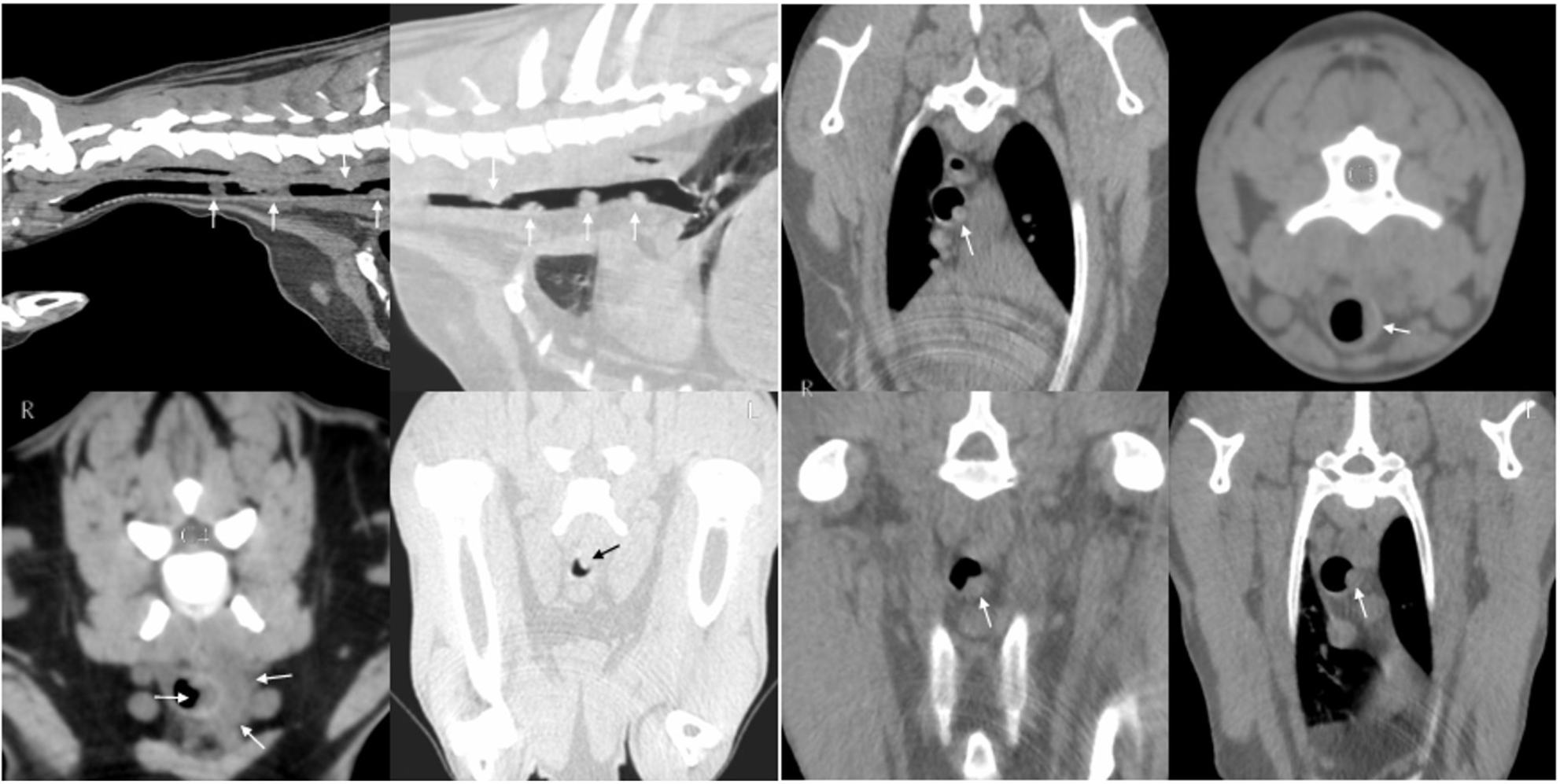
Advanced imaging demonstrated multifocal, nodular, eccentric thickening of the cervical and thoracic tracheal walls, predominantly involving the mucosa with extension along the adventitia, most severe at the level of C4.
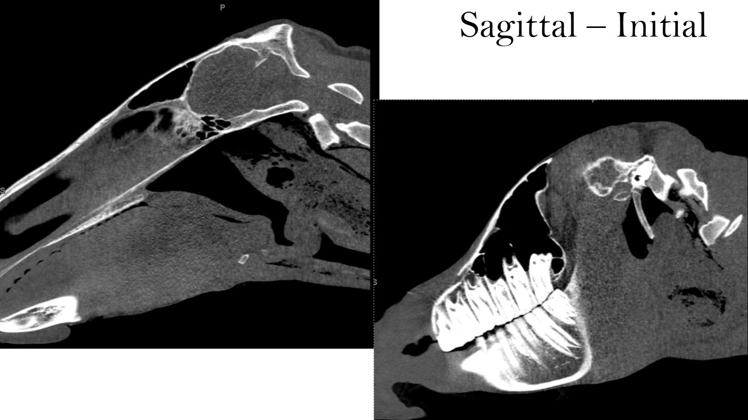
CT imaging demonstrated extensive peri-laryngeal and retropharyngeal subcutaneous emphysema with extravasated feed material and/or abscessation and regional cellulitis, consistent with suspected pharyngeal or cranial esophageal trauma or tear.
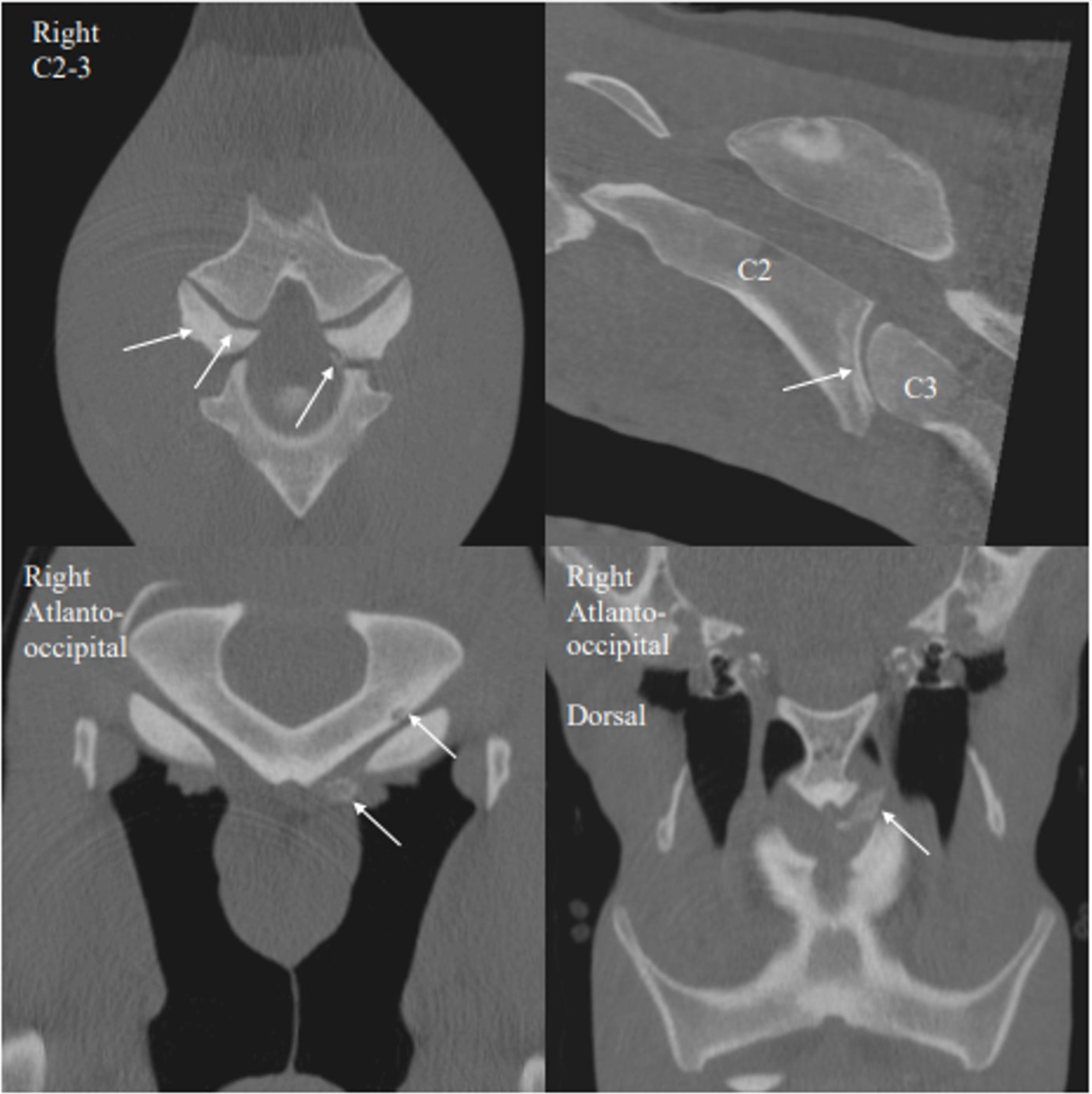
Advanced imaging identified moderate osteoarthritis of the C2–C3 articular process joints, more pronounced on the left, with a small axial mineral body/mineralization adjacent to the joint. Intervertebral disc disease was also present at C2–C3, without observable alteration in spinal cord position on this study.
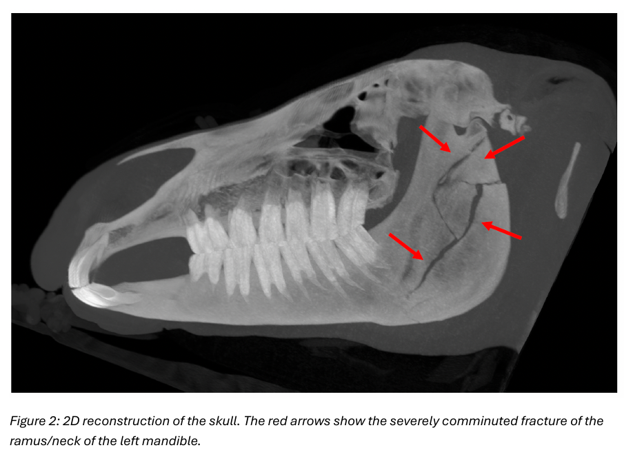
Severely comminuted, acute, mildly displaced closed and non-articular fracture of the ramus and neck of the left mandible. No evidence of TMJ luxation or joint incongruency was identified.
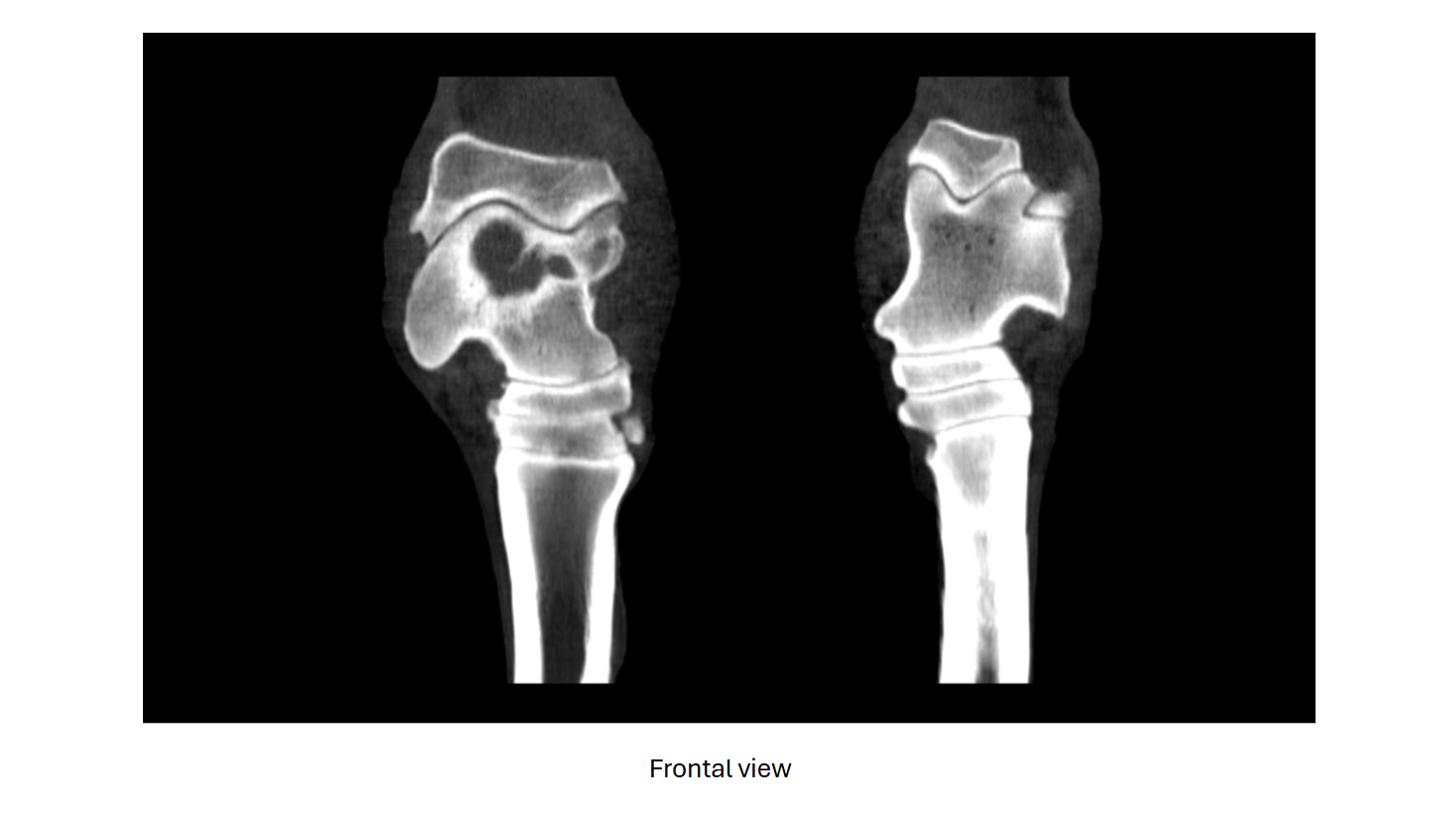
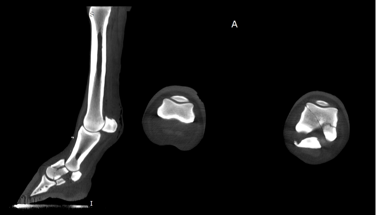
Advanced imaging revealed moderate tarsocrural joint osteoarthrosis and a large talar cyst with articular communication and an associated depression fracture, findings most consistent with a traumatic cyst.
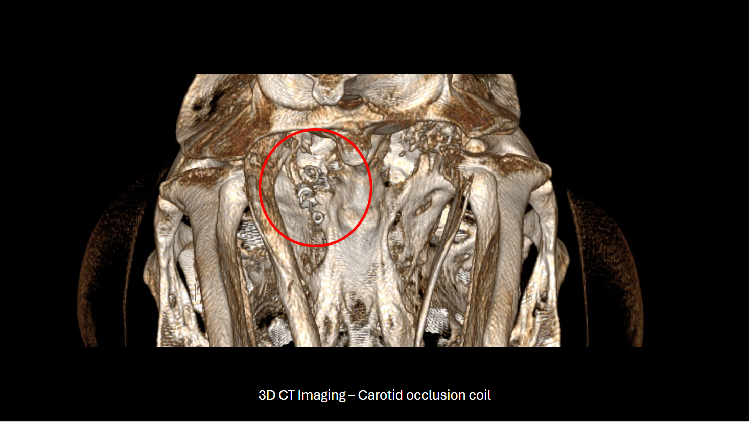
CT imaging demonstrated extensive occipital bone lysis and hyperostosis, regional infiltrative hypoattenuating soft tissue causing spinal cord compression, and marked thickening of the left stylohyoid bone and temporohyoid articulation

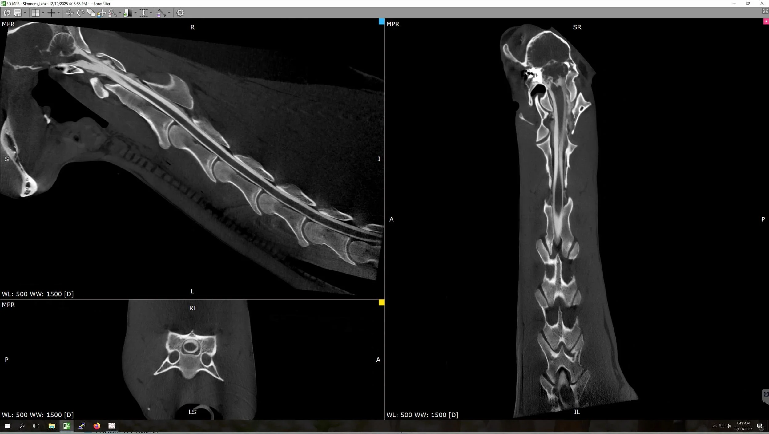
We're pleased to announce significant progress in deploying our technology to image the cervical spine. We were able to reach the cranial aspect of C7 at Virginia Equine Imaging on several standing patients.

3 yr old Gypsy Vanner gelding was referred for evaluation of a persistent left facial swelling that had been present for five months.

5 yr old TB gelding presented following an acute episode of severe pain, during which he went down and thrashed.


8 yr old warmblood gelding, concern for lateral collateral ligament involvement or possible OCD in the left hock.

A 13-year-old Gypsy mare was brought in with a several-month history of a protuberance from the right maxilla, which had recently started to drain. Radiographs indicated…

5-year-old Quarter Horse mare History: Presented with bilateral forelimb lameness; previous diagnoses include navicular syndrome and left front navicular bone cyst.

Patient was referred for evaluation of the head and neck after the referring veterinarian identified an abnormal noise and palpable movement in the cranial cervical region.
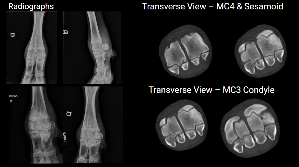
The CT identified osteochondritis dissecans, including a cystic lesion in the medial proximal sesamoid bone, lytic lesions in MC4, and subchondral defects in MC3.

5-year-old POA mare presented at the University of Minnesota for CT of the right front foot. She has an approximately 1-year duration of Grade 2/5 right front lameness.

9-month-old Percheron gelding presented for respiratory noise, leading to the discovery of a multilocular aneurysmal bone cyst.


16 year old Argentinian Warmblood Gelding presenting at the University of Minnesota for 4/5 right front lameness for a 10-14 days duration after 2 incidences of trauma (fighting with other horses).

24yo Arabian cross mare presented for unilateral left nasal discharge unresponsive to antibiotics.


Featured Images





Stay Connected
Subscribe to our Newsletter – Get the latest updates and exclusive offers delivered straight to your inbox.
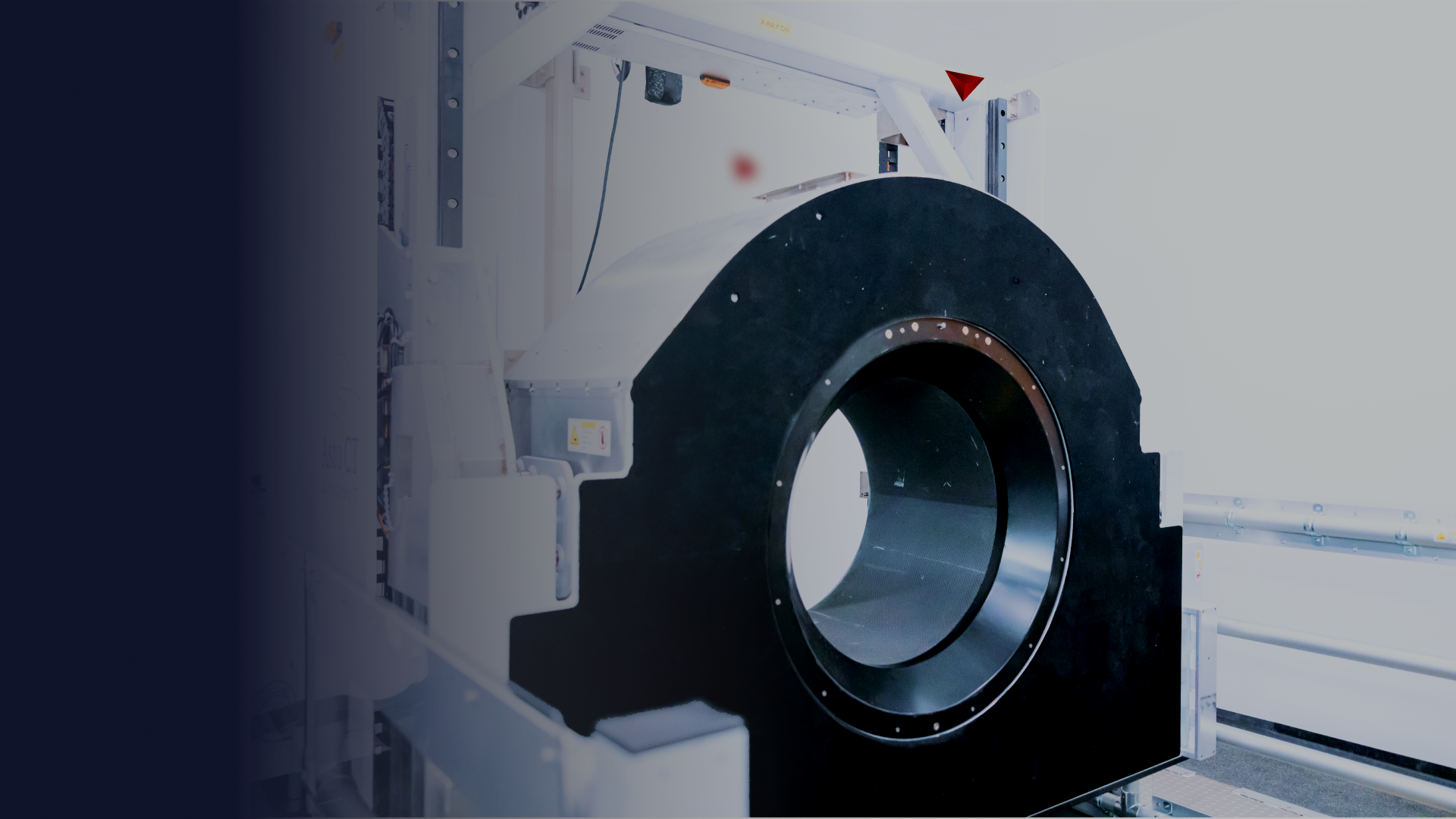
A new tool, Equina® (Asto CT, Middleton, WI), has been developed and specifically designed for a veterinarian to safely image a sedated standing horse in a natural load-bearing position solving some of the key barriers to equine CT imaging. In this paper, we provide a brief description of the key features of the Equina® system and describe how it can move us into a new equine standard-of-care.