
What Standing CT Is Teaching Us About Equine Diagnosis
May 2026 - Peer Reviewed
Standing computed tomography has not transformed equine practice because it is new technology, but because it has changed how, when, and how often advanced imaging is used. The clinical impact of standing CT lies not in its technicality, but in its accessibility and routine application. As barriers to use have fallen, clinicians are discovering that the pathology present in many horses is more extensive, more complex, and more multifocal than previously appreciated with radiography alone (Brounts et al 2022a).
The more relevant question today is no longer whether standing CT improves visualization, but instead what pathologies we are now able to diagnose and how this informs treatment. Given its widespread integration into comprehensive lameness evaluations and diagnostic workups, particularly for distal limb (Brounts et al 2022a) and diseases of the head and neck (Brounts et al b), it is advancing both the accuracy of diagnosis and the effectiveness of targeted treatment.
More Access Leads to More Answers
Experience in a University Setting
Over the past two and a half years at the University of Missouri College of Veterinary Medicine, more than 500 standing CT examinations have been performed in horses, the majority focused on the limbs. This level of utilization would have been inconceivable two decades ago. While equine limb CT has been technically possible for over 20 years, its reliance on general anesthesia restricted its use to a handful of cases annually. At most referral centers, two to three limb CT scans per year were typical.
The elimination of anesthetic risk has fundamentally changed that pattern. Equine clinics now perform well over 200 standing limb CT examinations annually. Scan times are shorter than MRI, clinicians get a larger area of interest per scan, and the modality is practical for routine use rather than reserved for complex cases.
Private Practice Setting
Similar trends have emerged in private practice imaging centers. Even in facilities with established access to nuclear scintigraphy, MRI, and ultrasonography, standing CT has rapidly demonstrated clinical value. Rather than utilizing existing modalities, it has filled a diagnostic gap between blocking and definitive diagnosis, particularly when lameness localization is challenging or when multiple joints warrant evaluation. Private practices leveraging a collaborative referral model to optimize internal and external referrals are seeing annual CT case volumes between 200-300.
In contrast to MRI examinations, which are time-intensive and typically scheduled well in advance, standing CT can be performed within minutes as part of a routine lameness evaluation. In both referred and in-house caseloads, Virginia Equine Imaging has evaluated approximately 350 regions of interest within the first year of installing a standing fan-beam CT system. These early outcomes highlight its clinical utility, with soft tissue findings supporting its role as a cost-effective and accessible alternative for select imaging indications traditionally addressed with MRI.
Commonly Seen with Standing Equine CT
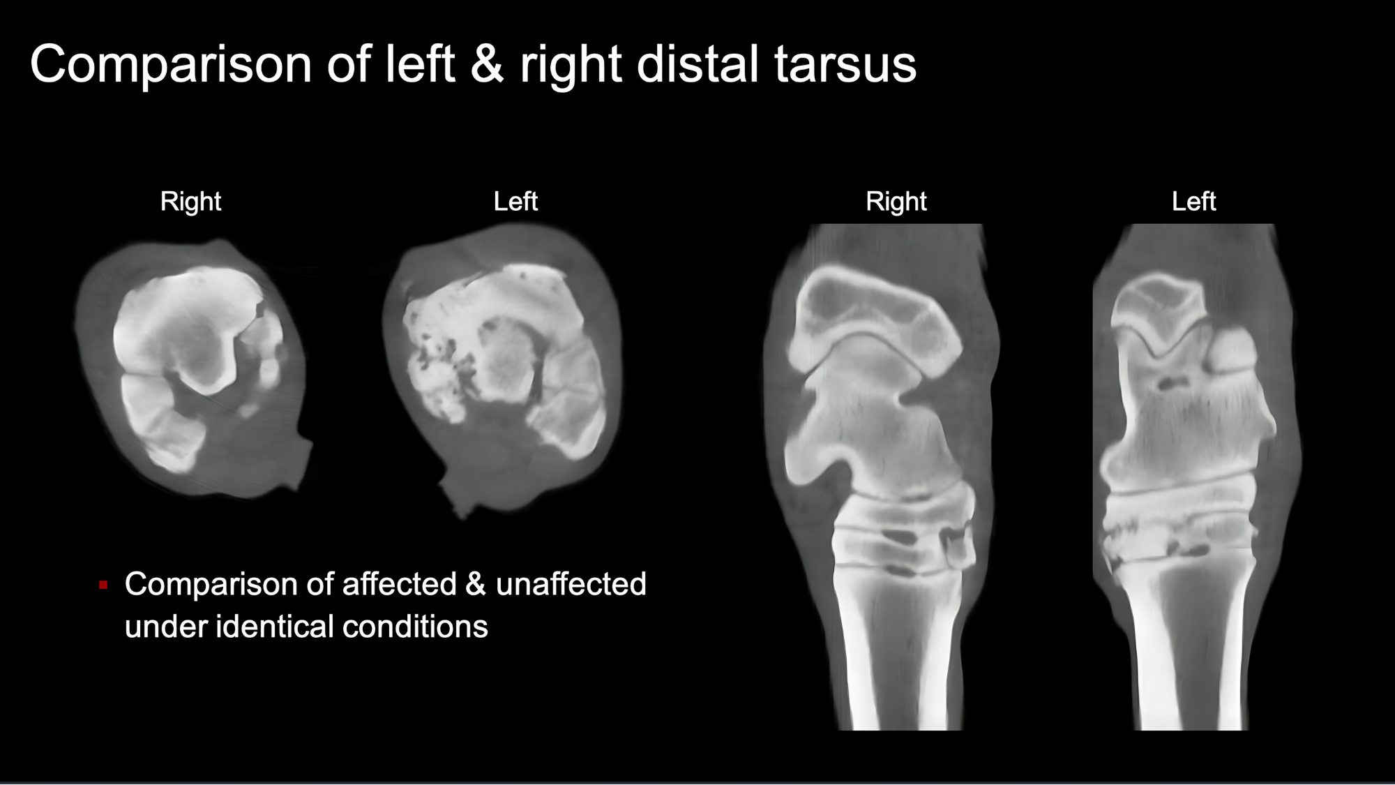
Figure 1
Distal Tarsal Joint Osteoarthritis
One of the most frequently identified conditions on standing CT is distal tarsal joint osteoarthritis (figure 1). While radiographs remain valuable, CT routinely reveals a broader and more severe spectrum of osseous change, including:
Extensive subchondral sclerosis
Focal or diffuse bone lysis
Small subchondral cystic lesions
These findings often explain persistent lameness that appears disproportionate to radiographic change and allow clinicians to better appreciate true disease burden.
“Seeing the combination of sclerosis, lysis, and cystic change together has completely changed how I assess distal hock OA,” says Dr. Megan McKraken DVM, MS, DACVS-LA.
Previously Under‑Recognized Sites
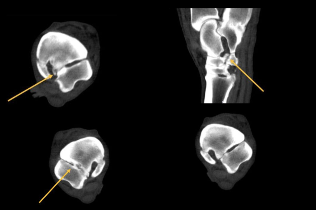
Figure 2
Standing CT has also shifted where clinicians look for pathology. Osteoarthritis between the central tarsal bone and the fourth tarsal bone (figure 2), for example, is increasingly recognized. This region is difficult to evaluate radiographically due to superimposition, yet pathology there is readily apparent on CT. As a result, disease previously overlooked or dismissed have become a routine part of diagnostic consideration.
In private practice, hind limb lameness suspected in the hock or proximal suspensory region is often difficult to define. Radiographs, diagnostic blocks, and ultrasound are useful, but some cases remain inconclusive. Standing CT adds value by providing detailed 3-D visualization of the plantar hock. It detects small cystic lesions and joint abnormalities not visible on radiographs. It also improves assessment of the proximal suspensory region, identifying bony proliferation, bone lysis, and subtle soft tissue lesions, including small ligament tears.
Subtle, Multifocal Bone Change
Perhaps one of the most clinically valuable insights from standing CT is how commonly lameness reflects subtle, multifocal bone change. Multiple joints are often involved, helping explain clinical signs that do not align with a single radiographic abnormality. This broader perspective has practical implications for both prognosis and treatment planning.
Cervical Spine and Neck Pathology
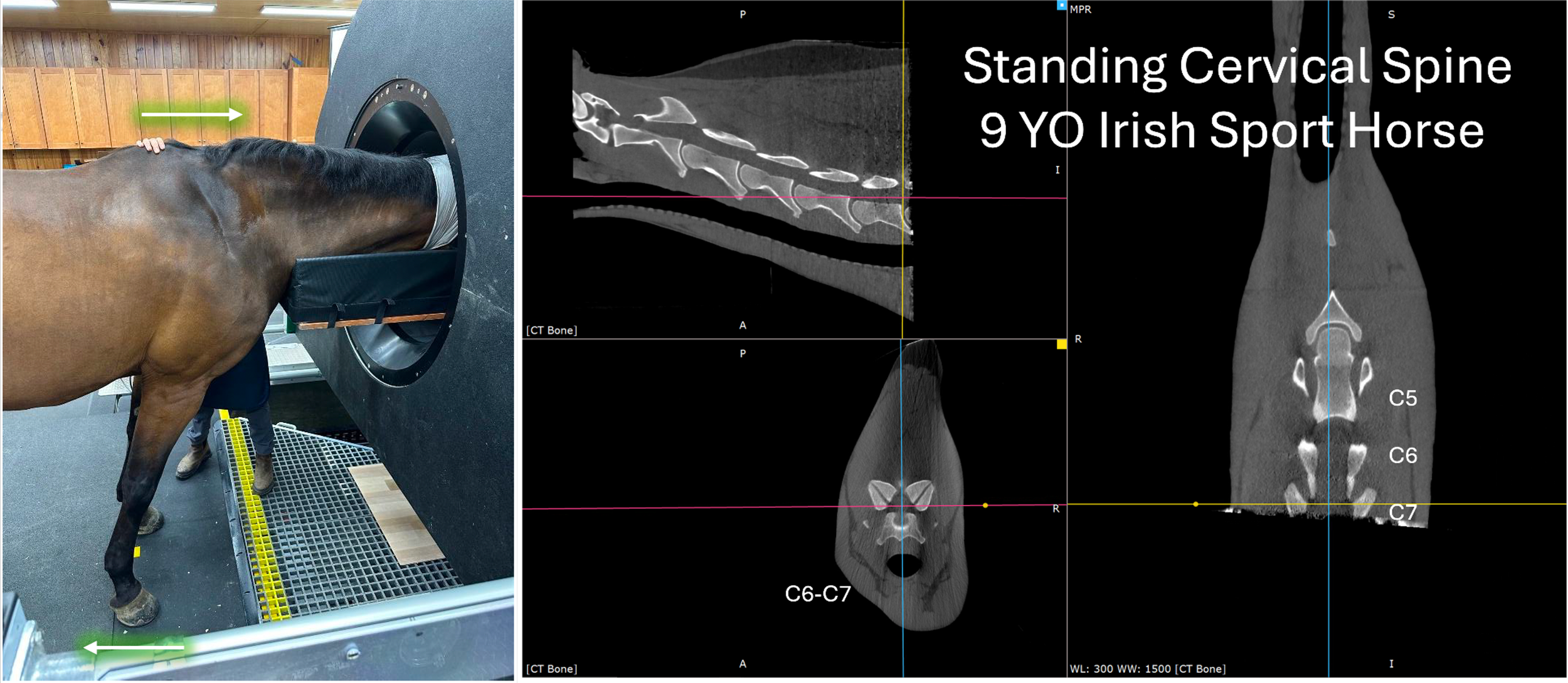
Figure 3
Standing CT has also expanded diagnostic capability beyond the limbs. Cervical spine imaging now routinely reaches C6–C7 standing in approximately 80–85 percent of horses (figure 3). Limitations relate more to positioning and patient compliance than to size or conformation, opening new diagnostic avenues for neurologic cases.
Dr. Jack Caldwell and the team at Virginia Equine Imaging developed a guide “Scanning the Complete Cervical Spine in the Standing, Sedated Horse: Tips and Tricks from Virginia Equine Imaging” outlining best practices for use of the Asto CT. It focuses on patient positioning, compliance, and safety to optimize cervical spine imaging results. Download the Guide Here
Head, Sinus, and Dental Disease
Common diagnoses include:
Periapical abscesses
Periodontal disease
Unilateral sinusitis
Skull fractures
In these cases, CT eliminates much of the ambiguity inherent in skull radiographs and is essential for surgical planning and postoperative assessment. In the first year following installation of the fan beam CT at Virginia Equine Imaging, head CT studies accounted for over 25% of the caseload, with an 89% positive diagnostic yield. At a recent continuing education event, Dr. Caldwell emphasized the advantages of fan-beam CT over radiography for accurate diagnosis of skull pathology.
The Impact of Earlier, More Accurate Diagnosis
The most impactful shift associated with standing CT is not simply improved image quality, but its placement earlier in the diagnostic pathway. Advanced imaging is no longer a final step undertaken after repeated examinations and treatments. Earlier diagnosis allows clinicians to implement targeted therapy sooner, avoiding trial‑and‑error methods and preserving long‑term musculoskeletal health.
CT does not guarantee a diagnosis. However, it consistently improves diagnostic confidence and narrows differentials, allowing for more informed decision‑making and better alignment between clinical findings and imaging results.
“Early, accurate diagnosis saves owners money, though this benefit is often underappreciated. By reducing repeated lameness evaluations and avoiding trial‑and‑error joint injections, we can implement targeted treatment sooner, allowing horses to return to comfort faster and maintain it for longer.”— Dr. Megan McCraken DVM, MS, DACVS-LA.
Cost, Practicality, and Clinical Value
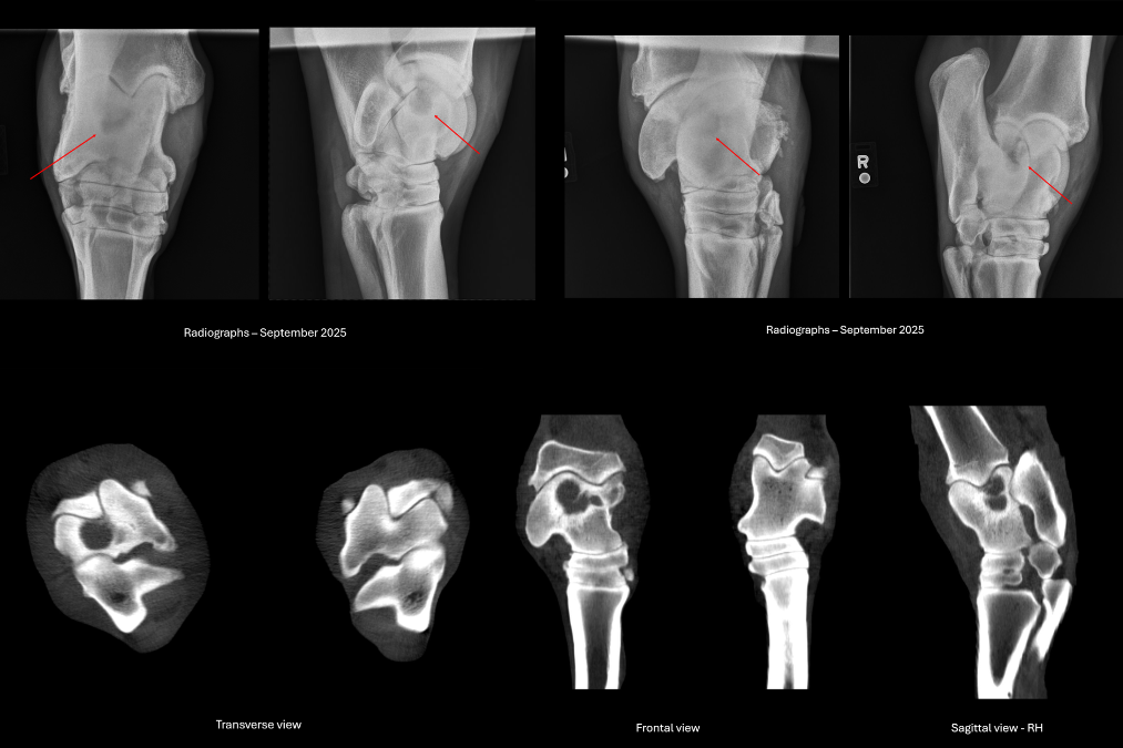
Figure 4: Right hind limb joint osteoarthrosis. Radiographs (top) demonstrate subtle lucencies that are more clearly and extensively appreciated on CT imaging (bottom).
As CT technology has advanced, costs have decreased and accessibility has increased. In many academic and private settings, bilateral weight‑bearing CT imaging from the tarsus or carpus to the foot costs approximately $370 more than bilateral hock radiographs (figure 4). When compared with the cumulative cost of multiple joint radiographic series, nuclear scintigraphy, or MRI, CT is often less expensive while providing a larger anatomic coverage area.
Standing CT is particularly valuable in horses that are difficult to block or in cases where blocking does not yield clear localization. Importantly, early accurate diagnosis frequently reduces overall expense by eliminating repeated lameness evaluations, unnecessary joint injections, prolonged referrals, or extended hospitalization. Horses are treated more precisely, return to comfort sooner, and maintain that comfort longer.
Education and Communication
Teaching Better Anatomy and Pathology
Standing CT has transformed veterinary education. Visualizing anatomy in multiple planes with three-dimensional reconstruction enables students to gain a more complete understanding of complex anatomic regions such as the tarsus and carpus.
Helping Owners Understand the Diagnosis
What may appear as a subtle greyscale change on radiographs becomes immediately understandable on CT. This clarity improves owner confidence in both diagnosis and treatment plans, strengthening veterinarian‑client communication.
Standing CT: an Important Tool in Advanced Lameness Practices
From an advanced imaging perspective, standing CT bridges the gap between blocking, diagnosis, and definitive treatment planning.
As Dr. Kent Allen, DVM, Virginia Equine Imaging, President of ISELP, observes:
“It makes sense that any equine practice with an advanced lameness program would have Equine Standing CT installed. For us at Virginia Equine Imaging, it’s a critical tool for diagnosing lameness and developing treatment plans. We’re seeing more detail, reaching diagnoses faster, and returning horses to their owners with clear answers. Horses with a diagnostic treatment plan can return to work sooner because we get answers faster.”
Customer outcomes show that clinics integrating Asto CT into an advanced lameness program can offset system costs through improved diagnostic efficiency. A typical referral clinic uses standing CT to replace or reduce repeated radiographs and anesthesia-based imaging. By shortening the diagnostic pathway and providing clearer early answers, clinics often see higher client acceptance rates and faster case resolution.
Conclusion
Routine use of standing CT has reshaped what equine veterinarians are diagnosing most often. Clinical experience has demonstrated that pathology is frequently more extensive, more advanced, and more multifocal than radiographs suggest. Standing CT accelerates confident decision‑making, improves diagnostic accuracy, and supports earlier, more targeted intervention. Fan-beam CT can equal or in some cases exceed MRI in diagnosing soft tissue injuries.
The conclusion is increasingly clear. Standing CT is no longer a last‑step diagnostic tool. Whether installed within a practice or accessed through a referral center, it fits seamlessly into the clinical work-up in case evaluation. Standing CT is not simply advanced imaging; it is better clinical reasoning, earlier intervention, and improved long‑term outcomes for horses.
References
Brounts, S.H., Lund, J.R., Whitton, R.C., Ergun, D.L., Muir, P. Use of a novel helical fan beam imaging system for computed tomography of the distal limb in sedated standing horses: 167 cases (2019-2020). J Am Vet Med Assoc. 2022a;260:1351-1360.
Brounts, S.H., Henry, T., Lund, J.R., Whitton, R.C., Ergun, D.L., Muir, P. Use of a novel helical fan beam imaging system for computed tomography of the head and neck in sedated standing horses: 120 cases (2019-2020). J Am Vet Med Assoc. 2022b;260:1361-1368.