C2–C3 Articular Process Joint Osteoarthritis
FEATURED WINNER – Neck Category
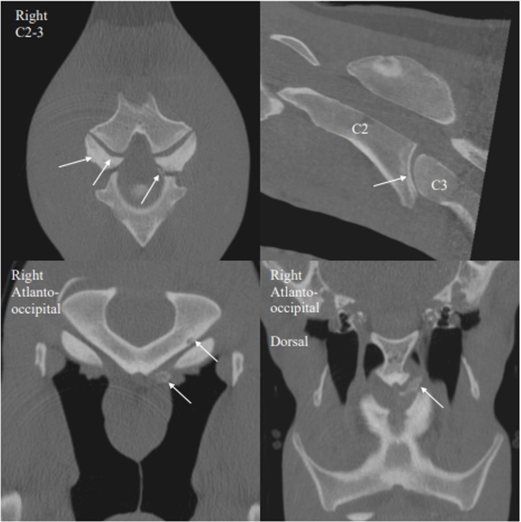
C2–C3 Articular Process Joint Osteoarthritis
6-year-old Quarter Horse gelding
University of Minnesota – Leatherdale Equine Center
Dr. Nicolas Ernst
Recognized for exceptional anatomical coverage and image quality, this case demonstrated CT’s value in diagnosing cervical pathology and assessing articular process joint disease with clarity beyond conventional imaging.
Case Summary
A 6-year-old Quarter Horse gelding was referred for advanced imaging due to head shaking and subtle facial asymmetry. At rest, the horse demonstrated mild deviation of the muzzle to the left. When stressed, a twitch originating at the left nostril and extending across the face to the left ear was observed. No nasal discharge was reported, though the gelding resisted turning to the left during barrel racing maneuvers, raising concern for cervical or neurologic involvement.
Diagnosis
Advanced imaging identified moderate osteoarthritis of the C2–C3 articular process joints, more pronounced on the left, with a small axial mineral body/mineralization adjacent to the joint. Intervertebral disc disease was also present at C2–C3, without observable alteration in spinal cord position on this study. Additional findings included a small subchondral bone cyst–like lesion of the left occipital condyle and a small osseous body ventral to the condyle. These changes were considered potentially developmental, traumatic, degenerative, or multifactorial in origin, with questionable clinical relevance. The C2–C3 degenerative changes were considered the most likely contributors to the gelding’s reported performance-related issues.