
Whitepaper: A Novel High-Quality Equine Standing CT Scanner: The Asto CT Equina
David Ergun 1,3, PhD, CEO
Sabrina Brounts 1,2, DVM, MS, PhD, DACVS/ECVS, DACVSMR, Scientific Advisor
Rock Mackie 1,3, PhD, Chief Innovation Officer
1: Asto CT Inc, Middleton, WI USA
2: School of Veterinary Medicine, University of Wisconsin-Madison, Madison, WI USA
3: Medical Physics, University of Wisconsin-Madison, Madison, WI USA
Revised May 2026
Introduction
With hundreds of thousands of horses used for racing, sport, and recreation worldwide, the effective management of injury and disease throughout a horse’s lifetime remains a central priority in equine veterinary care. Although imaging is a critical component of diagnosis and treatment planning, it is often underutilized despite its potential to significantly improve clinical outcomes.
Widely available and cost-effective modalities such as radiography and ultrasound often lack sufficient diagnostic sensitivity, particularly for early or complex pathology. In contrast, advanced volumetric imaging technologies such as computed tomography (CT) and magnetic resonance imaging (MRI) have historically required general anesthesia and complex positioning, limiting their practicality in routine clinical settings. In addition, some systems are not specifically designed for equine patients, resulting in reduced reliability and suboptimal image quality.
The Equina® system (Asto CT Inc, Middleton, WI) is a novel CT platform specifically designed to enable safe and effective imaging of the sedated standing horse in a natural weight-bearing position. By addressing a key barrier to equine CT imaging, the system expands access to advanced diagnostic imaging in equine practice. In this paper, we describe the key features of the Equina® system and its potential to establish a new standard of care.
How does Equina® work?
The Equina® is a dual-direction CT scanner that can be used to image a sedated, standing horse. The critical innovation is the integration of flexible positioning capabilities with a clinical-grade CT gantry. This design allows the gantry to be moved into the appropriate position for imaging the limbs or the head and neck without requiring complex positioning of the horse or the use of general anesthesia. During image acquisition, the scanner moves while the patient remains stationary.
Image acquisition and reconstruction use a state-of-the-art helical fan beam CT scanner powered by a 240V single-phase 30A uninterruptible power supply (UPS). Scanning is performed at 160 kVp with a rotation time of 1 second per revolution. The system includes 24 detector rows with a helical pitch of 0.55. The slice acquisition rate is 36 slices/sec with an image matrix of 1024 x 1024 and a spatial resolution of 0.75 mm at isocenter.
The CT gantry has a bore diameter of 75 cm with a field of view (FOV) of 75 cm, meaning that everything within the bore opening is imaged. This is an important design feature, as it greatly simplifies patient positioning for both limb and head scans of the standing horse and eliminates the need for a scout scan. The maximum scan distance is 75 cm for limb imaging and 92 cm for head and neck imaging, both at a translation speed of 2 cm/sec.
The operation and movement of the CT scanner are controlled by a simple hand-held controller with push-button controls, allowing the operator to remain close to the patient and handlers during scanning. For patient safety, a release switch on the system pendant controller can be activated to quickly return the CT gantry to its home position beneath the platform.
The inherently low power of the CT scanner, combined with a high level of internal self-shielding, allows patient handlers wearing standard protective shielding (apron, thyroid, and eye protection) to remain in the room during scanning, thereby optimizing patient handling and safety (Veitch et al. 2025).
For limb scanning (Figure 1), the CT system images either the two thoracic (front) limbs or the two pelvic (rear) limbs in a natural load-bearing position. As shown in Figure 1A, the CT gantry is initially positioned below the system platform. The sedated horse is then walked across the platform so that the limb pairs are positioned within the bore of the gantry (Figure 1B). The operator raises the gantry to the desired start position and initiates the scan. Scans typically take less than 20 seconds, after which the gantry returns below the platform and the horse can be safely removed.
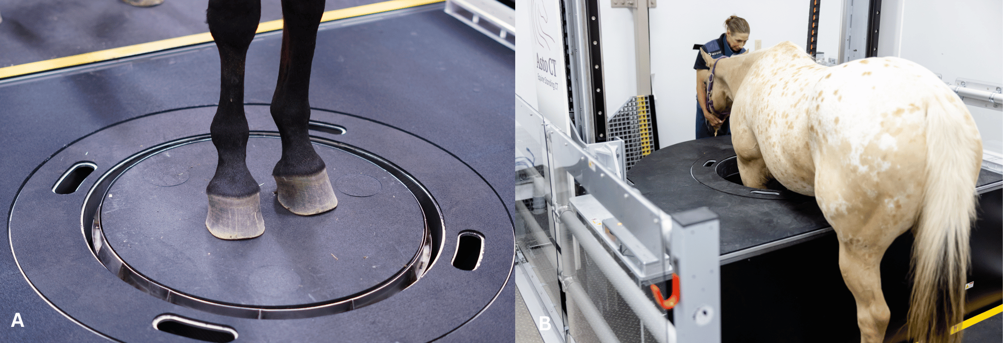
Figure 1. Set-up for a CT scan of distal thoracic limbs in a horse. The CT gantry sits below the system platform (A).
The limb pairs are positioned with the bore of the CT gantry for scanning (B). (Photo Courtesy of Wisconsin Equine Clinic & Hospital)
For head and neck imaging (Figure 2), the CT gantry is raised and rotated to allow horizontal scanning (Figure 2A). The horse is positioned in stocks with a cushioned headrest (Figure 2B). The gantry is adjusted to a comfortable height, and scanning is performed as the gantry advances along the head and neck. Scan times can be up to 45 seconds depending on the region of interest.
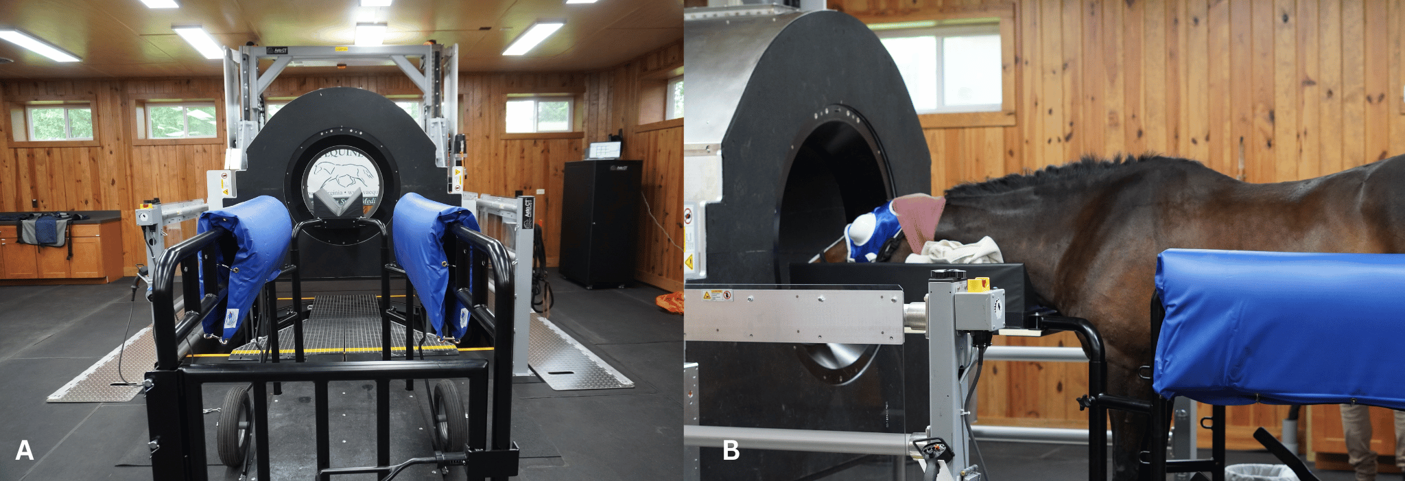
Figure 2. Set-up for a CT scan of the head and neck region in a horse. The CT gantry is raised from the floor and rotated
into a vertical position for horizontal scanning (A). The sedated horse stands in the stocks with its head on a headrest (B). (Courtesy of Virginia Equine Imaging.)
Using the standard stock configuration, imaging can extend to approximately the C4–C5 level. For more caudal imaging, a rear-mounted cushioned headboard can be used (Figure 3), allowing extension to C6–C7. As illustrated in Figure 3 (A and B), this accessory enables the horse to lean forward comfortably into the bore, facilitating imaging of the caudal cervical region.
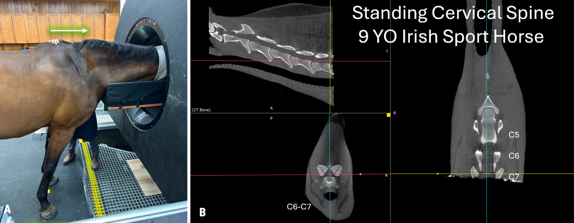
Figure 3: Setup for CT imaging of the caudal cervical region using a rear-mounted headboard. The headboard is positioned at the rear of the scanner and extends forward into the bore (A), allowing the sedated horse to lean forward and enabling imaging to C6-C7 (B). (Courtesy of Virginia Equine Imaging.)
While the primary value of the Equina® system is standing imaging, there are cases where imaging under general anesthesia is desirable, such as in horses with neurological conditions. In these situations, a surgical table extension can be used to position the patient in dorsal or lateral recumbency (Figure 4). This configuration allows imaging of regions such as the cervical spine under general anesthesia, including procedures such as myelography (Figure 5).
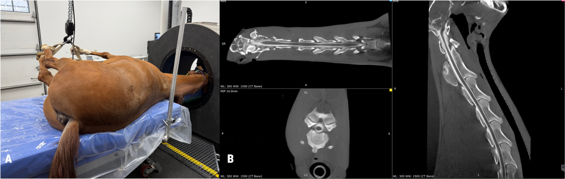
Figure 4: Setup for CT imaging of the cervical region in a horse under general anesthesia. The horse is positioned in lateral recumbency on a surgical table, with the CT gantry in the home position. (Courtesy of Wisconsin Equine Hospital & Clinic.)
EQUINA® IN CLINICAL PRACTICE – LIMBS
Lameness is a common problem in horses and can be challenging to diagnose. Diagnostic imaging plays a central role in identifying the cause. While radiography and ultrasound are widely used, early detection of subtle or complex pathology can be challenging. Standing CT provides high-resolution imaging of both bone and soft tissue, improving diagnostic accuracy.
Equina® has been used to assess the limbs of several thousand equine patients since 2018. In a study of 167 horses, 88 showed diagnosable findings on CT (Brounts et al. 2022a). The most common findings included navicular syndrome in the foot/pastern region and palmar/plantar osteochondral disease in the fetlock. In cases where both radiographs and CT were performed, only 25% of diagnoses were identified using radiography alone, indicating a substantial limitation of conventional imaging.
Importantly, no complications were reported, and no equipment failures occurred. These findings support the safety and clinical value of the Equina® system for limb imaging.
EQUINA® IN CLINICAL PRACTICE – HEAD and neck
The Equina® system also enables efficient imaging of the head and neck. Radiographic evaluation of the head is often limited by anatomical complexity and superimposition. CT imaging provides clear cross-sectional detail, improving diagnostic confidence.
A study of 120 horses undergoing head and neck CT imaging demonstrated higher diagnostic efficacy compared to radiography (Brounts et al. 2022b). The most common findings included dental disease and sinus lesions, while neck imaging identified conditions such as nuchal bursitis and spinal abnormalities. No complications were reported, reinforcing the safety of the system.
THE UNIQUE FEATURES OF THE EQUINA®
The Equina® system was purpose-built in collaboration with equine veterinarians, resulting in an integrated design that prioritizes safety, durability, and clinical usability across a wide range of patient sizes and conditions. Key design features include:
A laser curtain that prevents the gantry from contacting the patient during movement.
A rugged, non-slip, shock-absorbing gantry surface that allows horses to safely walk across the system.
A sealed gantry and motion system that protects against dust, water, and other contaminants, with surfaces designed for easy cleaning.
The physical geometry of the system further enhances its versatility. For head and neck imaging, the bore opening measures 92 cm at the entrance and tapers to 75 cm internally, facilitating positioning for cervical spine imaging. This configuration supports routine standing imaging of the neck to the level of C7 when used with the rear-mounted headboard (Figure 5).
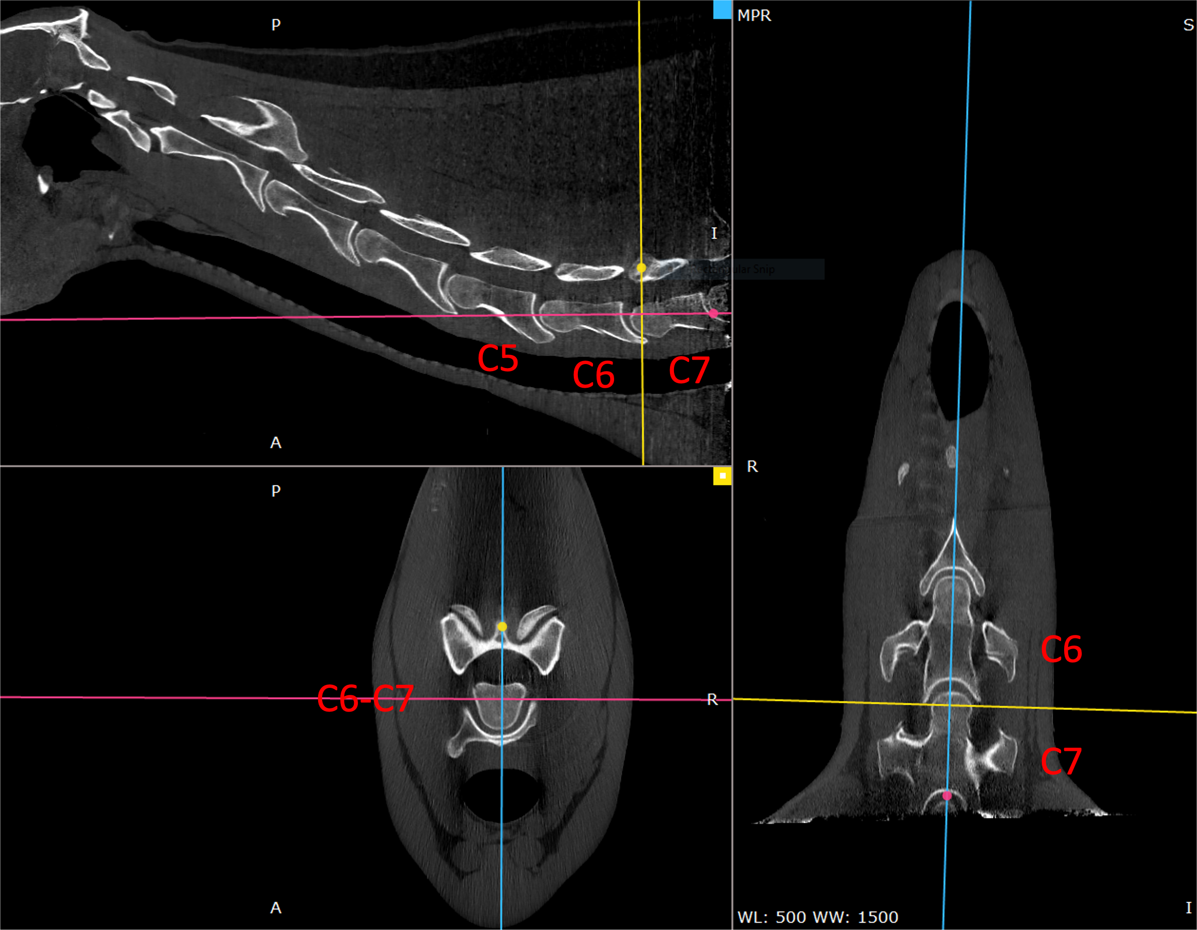
Figure 5: Representative images from a standing neck CT scan demonstrating cervical imaging to the level of C7. Images include AI-enhancement for improved visualization.
Imaging performance is optimized through protocols tailored specifically for equine anatomy. Operation at 160 kVp provides excellent visualization of dense subchondral bone, while dedicated reconstruction filters enable detailed assessment of both bone and soft tissue structures. Full-field reconstructions are available immediately following acquisition (Figure 6).
Finally, the system’s low power requirements and integrated self-shielding allow personnel to remain in the room during scanning without exceeding recommended occupational exposure limits when appropriate protective equipment is used (Veitch et al. 2025). This contributes not only to safety but also to improved workflow and patient handling.
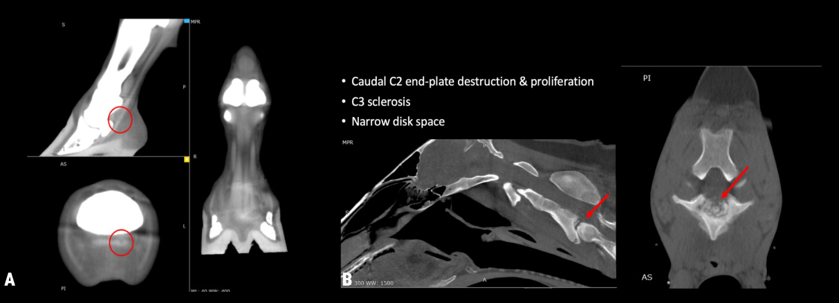
Figure 6: CT dataset reconstructed using a soft-tissue filter in a 13-yr-old Quarter Horse with tendon lesion, demonstrating soft-tissue contrast (A). CT dataset reconstructed using a bone filter in a 2-year-old Morgan with a neurologic disorder, highlighting bone detail (B).
TRANSFORMING THE STANDARD OF CARE
The Equina® system is currently in use in multiple clinical settings such as academia, referral centers, and private practices and has demonstrated its ability to enhance diagnostic capabilities in equine practice. By enabling fast, safe, and standing CT imaging, it provides veterinarians with a powerful tool to improve patient outcomes.
Applications include:
Pre-purchase examinations
Lameness evaluation and management
Monitoring for stress fractures in performance horses
Rehabilitation monitoring
Dental and sinus evaluation
Image-guided procedures
Surgical planning
Non-equine species such as ruminants (Means et al. 2026), camelids, and small animals
SUMMARY
Computed tomography has significantly advanced equine imaging, particularly for the head and orthopedic conditions. However, its use has traditionally been limited by the need for general anesthesia.
The Equina® system overcomes these limitations by enabling standing CT imaging with minimal stress to the horse. Its versatility, safety, and ease of use position it as a transformative technology that supports a higher standard of care in equine veterinary practice.
REFERENCES
Brounts SH, Lund JR, Whitton RC, Ergun DL, Muir P. Use of a novel helical fan beam imaging system for computed tomography of the distal limb in sedated standing horses: 167 cases (2019–2020). J Am Vet Med. 2022;260(11):1351-1360. doi.org/10.2460/javma.21.10.0439
Brounts SH, Henry T, Lund JR, Whitton RC, Ergun DL, Muir P. Use of a novel helical fan beam imaging system for computed tomography of the head and neck in sedated standing horses: 120 cases (2019–2020). J Am Vet Med. 2022;260(11): 1361-1368. doi.org/10.2460/javma.21.10.0471
Means, K. L., Loeber, S., & Brounts, S. H. (2026). Standing computed tomography is feasible and has clinical utility in ruminants. American Journal of Veterinary Research, 87(1), Article ajvr.25.07.0254, ajvr.25.07.0254. Retrieved Mar 13, 2026, from https://doi.org/10.2460/ajvr.25.07.0254
Veitch KE, Szczykutowicz TP, Brounts SH, Ergun DL, Muir P, Loeber SJ. Radiation exposure during simulated equine head and limb fan beam standing computed tomography appears safe for personnel using lead shielding. J Am Vet Med. 2025;263(1): 63-70. doi.org/10.2460/javma.24.06.0424
©2026 Asto CT Inc – All rights reserved. Asto CT and Equina are registered trademarks of Asto CT.
Dr. Ergun, Dr. Brounts and Dr. Mackie have financial interest in Asto CT