
Asto CT Honors 2025’s Best CT Cases: Showcasing Outstanding Equine Diagnostics
We are proud to announce the winners of the 2025 Asto CT Best CT Cases of the Year, recognizing exceptional clinical cases that highlight the diagnostic power, versatility, and clinical impact of standing CT imaging across equine and veterinary medicine.
This year’s submissions showcased outstanding image quality, innovative case selection, and meaningful contributions to patient diagnosis and management. We extend our sincere thanks to all clinicians who participated and continue to push the boundaries of advanced imaging.
Below are the first-place winners by category, representing excellence in 3D reconstruction, limb imaging, head and neck diagnostics, soft tissue evaluation, and non-equine applications.
3D CATEGORY-10 entries
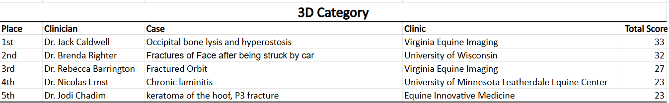
FEATURED WINNER – First Place
Occipital Bone Lysis and Hyperostosis
15-year-old Thoroughbred gelding
Virginia Equine Imaging
Dr. Jack Caldwell
Highest Scoring Entry with a score of 33 out of 35 points.
This case was recognized for its exceptional 3D reconstruction quality and its ability to clearly define complex bony pathology in the occipital region—information that would have been difficult to fully appreciate using traditional imaging alone.
Case Summary
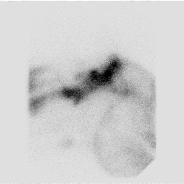
Nuclear scintigraphy
A 15-year-old Thoroughbred gelding was referred for full-body nuclear scintigraphy due to bilateral hindlimb lameness, decreased cervical range of motion, and Grade 2–3 ataxia. The gelding had a prior history of guttural pouch mycosis treated three years earlier with coil embolization of the left carotid artery. Nuclear scintigraphy revealed intense focal radiopharmaceutical uptake at the external occipital protuberance (poll) and C1, prompting advanced CT evaluation of the caudal head and cranial cervical spine.
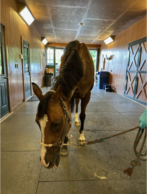
Clinical Signs
At rest, the gelding stood with his head lowered and deviated to the right.
Diagnosis
CT imaging demonstrated extensive occipital bone lysis and hyperostosis, regional infiltrative hypoattenuating soft tissue causing spinal cord compression, and marked thickening of the left stylohyoid bone and temporohyoid articulation. Findings were most consistent with fungal osteomyelitis and meningitis, given the history of guttural pouch mycosis.


Outcome & Post-Mortem Findings
Due to the poor prognosis associated with progressive neurologic deterioration and suspected fungal infection, euthanasia was elected. Post-mortem examination confirmed marked lysis of the occipital and atlas (C1) bones, Wallerian degeneration of the proximal cervical spinal cord, and cerebrospinal fluid culture positive for Aspergillus species, most closely resembling the A. fumigatus complex.


LIMBS CATEGORY-26 entries

Four-way tie between third place.
FEATURED WINNER – First Place
Tarsal Osteoarthrosis with Cyst
11-year-old Thoroughbred gelding
Virginia Equine Imaging
Dr. Jack Caldwell
Judges awarded high marks for the scan’s diagnostic clarity and clinical relevance. The CT study provided detailed evaluation of joint degeneration and cystic change, directly supporting diagnosis and case management.
Case Summary
An 11-year-old Thoroughbred gelding was referred for advanced imaging of the tarsus due to persistent right hindlimb lameness. In September 2024, the gelding sustained a fracture of the right lateral malleolus of the tibia during recovery from colic surgery. The fracture fragment was surgically removed one week later; however, intermittent right hindlimb lameness persisted, prompting further diagnostic evaluation.
Diagnosis
Right Hindlimb:
Advanced imaging revealed moderate tarsocrural joint osteoarthrosis and a large talar cyst with articular communication and an associated depression fracture, findings most consistent with a traumatic cyst. Additional findings included multifocal periosteal reaction consistent with regional inflammation and post-surgical change, lateral and medial collateral ligament enthesopathies, mild proximal interphalangeal joint osteoarthrosis, and moderate distal intertarsal joint osteoarthrosis.
Left Hindlimb:
No significant abnormalities were identified.


HEAD CATEGORY-19 entries
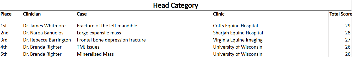
FEATURED WINNER – First Place
Fracture of the Left Mandible
15-year-old Welsh Section A mare
Cotts Equine Hospital
Dr. James Whitmore
This case stood out for its precise visualization of mandibular fracture configuration, offering critical insight into fracture extent and alignment that informed treatment planning.
Case Summary
A 15-year-old Welsh Section A mare was referred for advanced imaging following multiple traumatic kicks to the head, with concern for injury to the left mandible and temporomandibular joint (TMJ). Clinically, the mare was dull and exhibited abnormal left optic reflexes, left-sided facial nerve deficits (drooping ear and muzzle), hypersalivation with dysphagia, and increased respiratory effort with inspiratory stridor. Due to the severity of neurologic and upper airway signs, a standing head CT was performed.
Imaging Findings
Standing head CT performed on November 11, 2025, revealed a severely comminuted, acute, mildly displaced closed and non-articular fracture of the ramus and neck of the left mandible. No evidence of TMJ luxation or joint incongruency was identified. A small, acute, mildly displaced non-articular fracture of the left paracondylar process was also present.
A faint hypoattenuating line within the left parietal bone/zygomatic arch was noted and considered most consistent with a vascular channel, with fissure formation considered less likely. Marked axial and abaxial soft tissue swelling was present along the left side of the head, resulting in moderate to severe pharyngeal narrowing. The left guttural pouch was collapsed and/or filled with soft tissue attenuation material, consistent with hematoma formation or compression secondary to surrounding soft tissue swelling.


NECK CATEGORY-13 entries
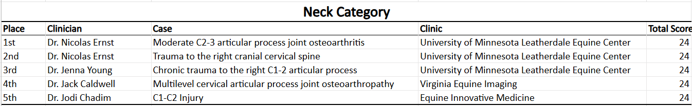
FEATURED WINNER – First Place
C2–C3 Articular Process Joint Osteoarthritis
6-year-old Quarter Horse gelding
University of Minnesota – Leatherdale Equine Center
Dr. Nicolas Ernst
Recognized for exceptional anatomical coverage and image quality, this case demonstrated CT’s value in diagnosing cervical pathology and assessing articular process joint disease with clarity beyond conventional imaging.
Case Summary
A 6-year-old Quarter Horse gelding was referred for advanced imaging due to head shaking and subtle facial asymmetry. At rest, the horse demonstrated mild deviation of the muzzle to the left. When stressed, a twitch originating at the left nostril and extending across the face to the left ear was observed. No nasal discharge was reported, though the gelding resisted turning to the left during barrel racing maneuvers, raising concern for cervical or neurologic involvement.
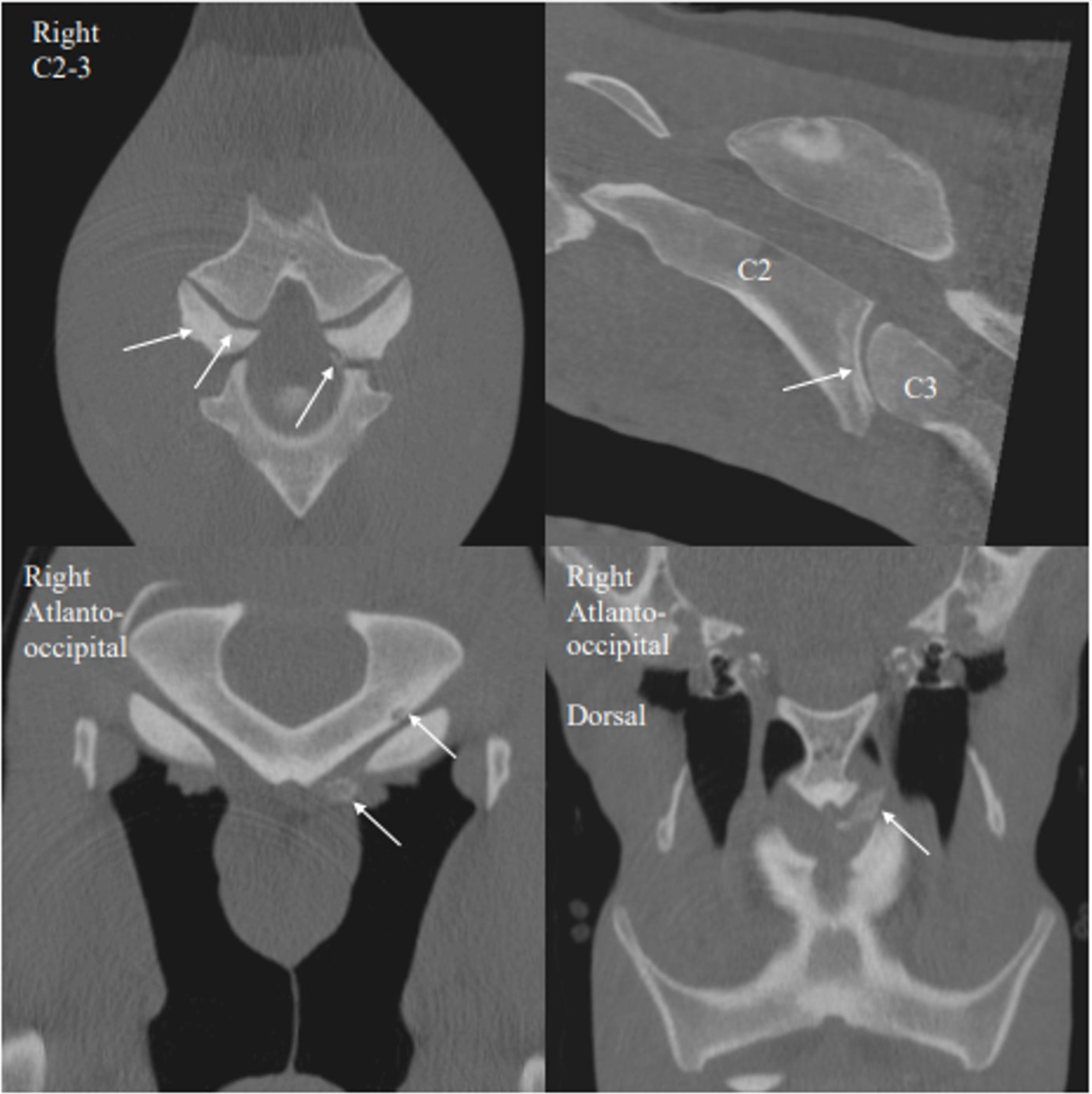
Diagnosis
Advanced imaging identified moderate osteoarthritis of the C2–C3 articular process joints, more pronounced on the left, with a small axial mineral body/mineralization adjacent to the joint. Intervertebral disc disease was also present at C2–C3, without observable alteration in spinal cord position on this study. Additional findings included a small subchondral bone cyst–like lesion of the left occipital condyle and a small osseous body ventral to the condyle. These changes were considered potentially developmental, traumatic, degenerative, or multifactorial in origin, with questionable clinical relevance. The C2–C3 degenerative changes were considered the most likely contributors to the gelding’s reported performance-related issues.
SOFT TISSUE CATEGORY-13 entries
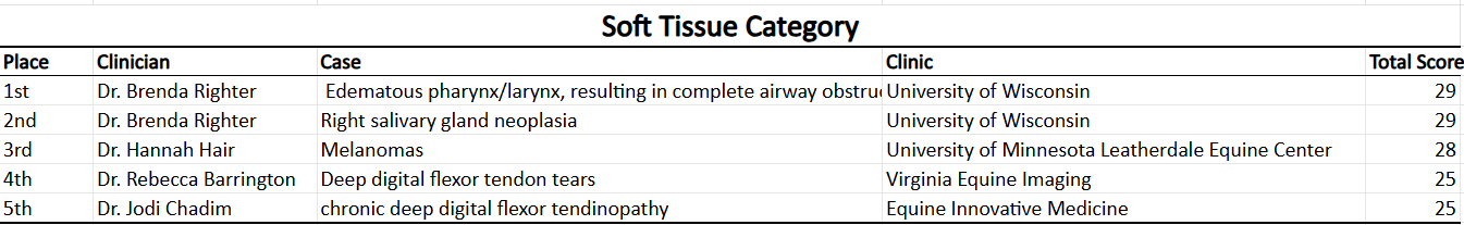
FEATURED WINNER – First Place
Peri-laryngeal Emphysema with Abscess
8-year-old Quarter Horse gelding
University of Wisconsin–Madison
Dr. Brenda Righter
This winning soft tissue case highlighted CT’s ability to evaluate complex airway and peri-laryngeal pathology, providing crucial diagnostic detail that significantly influenced clinical decision-making.
Case Summary
An 8-year-old Quarter Horse gelding (485 kg) was referred for evaluation of a suspected esophageal tear following hospitalization for colic at another facility. During the prior hospitalization, the horse received frequent nasogastric intubation for oral fluid administration and underwent multiple diagnostic procedures, including gastroscopy, upper airway endoscopy, proximal duodenal biopsy, oral glucose absorption testing, and dental floatation. Although colic signs resolved with medical management, the horse subsequently developed pharyngeal swelling with crepitus, dyspnea, fever, and hyporexia, prompting placement of a temporary tracheostomy prior to referral.
Clinical Presentation
On arrival, the gelding was quiet, hypersalivating, and grinding his teeth. Vital parameters were within normal limits. Diffuse subcutaneous emphysema with marked crepitus was present throughout the pharyngeal and proximal cervical regions, more severe on the left. Jugular refill time and cranial lymph nodes could not be assessed due to extensive swelling. Mucoserous secretions were observed at the tracheostomy site, and portions of the cervical skin felt cool on palpation.
Diagnostics & Imaging Findings
Upper respiratory and esophageal endoscopy revealed severe edema of the pharynx and larynx resulting in complete airway obstruction, with normal guttural pouches and no visualized esophageal or tracheal perforation. Due to concern for ongoing pathology and the possibility of healed mucosal injury, standing CT of the head and neck was performed. CT imaging demonstrated extensive peri-laryngeal and retropharyngeal subcutaneous emphysema with extravasated feed material and/or abscessation and regional cellulitis, consistent with suspected pharyngeal or cranial esophageal trauma or tear. Secondary findings included progressive laryngeal compression and laryngitis, ventral cervical emphysema tracking toward the vertebral canal, tracheitis and tracheal edema likely associated with the tracheostomy tube, over-riding tracheal membranes related to regional inflammation or trauma, and mild right rostral maxillary sinusitis.
Clinical Course & Outcome
High-dose penicillin therapy and supportive care resulted in marked improvement in subcutaneous emphysema and external swelling. Serial CT and endoscopic evaluations demonstrated progressive resolution of retropharyngeal abscessation, cellulitis, and emphysema, with improvement in arytenoid function and resolution of laryngitis. By late September, only minimal residual emphysema remained. The gelding continued to improve clinically, with normalization of attitude and appetite. Anti-inflammatory and antimicrobial therapies were gradually tapered, and the horse was discharged for continued home care with plans for follow-up upper airway endoscopic evaluation.


NON-EQUINE CATEGORY-10 entries
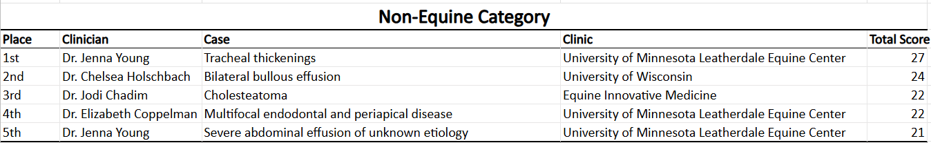
FEATURED WINNER – First Place
Thickening of the Cervical and Thoracic Tracheal Wall
4-year-old Nigerian Dwarf doe
University of Minnesota
Dr. Jenna Young
This submission exemplified the expanding applications of Asto CT technology beyond equine medicine, showcasing its diagnostic value in non-equine species and complex airway evaluation.
Case Summary
A 4-year-old Nigerian Dwarf doe was presented on an emergency basis for acute respiratory distress. Prior thoracic radiographs and ultrasound performed by the referring veterinarian revealed no significant abnormalities. On physical examination, the doe exhibited marked inspiratory stridor. Upper airway endoscopy identified irregular, proliferative tissue adhered to the tracheal wall extending from the mid trachea to the carina. The tissue was vascular and friable, occupying up to one-third of the tracheal lumen in certain regions.
Diagnosis
Advanced imaging demonstrated multifocal, nodular, eccentric thickening of the cervical and thoracic tracheal walls, predominantly involving the mucosa with extension along the adventitia, most severe at the level of C4. Secondary tracheal deformation and luminal narrowing were present, with up to 50% obstruction in affected areas, correlating with the patient’s clinical signs. Differential diagnoses included inflammatory or infectious processes such as granulomatous disease (parasitic, eosinophilic, abscessation, or papillomatosis) as well as neoplastic conditions including lymphoma or carcinoma.

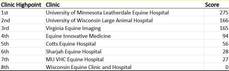
Overall Clinic Highpoint
91 entries total-31 entries counted towards highpoint
Congratulations to University of Minnesota Leatherdale Equine Hospital with a score of 275! The top 5 scores from all 6 categories were included in the cumulative results. UMN will pick out a custom lead apron from Philips Safety.
👏 Congratulations to all winners and thank you to every clinician who submitted a case this year. Your dedication and expertise continue to elevate the standard of care in veterinary medicine. Also thank you to our judges Dr. Kurt Selberg and Dr. Myra Barrett.
We look forward to seeing even more innovative and impactful cases in 2026.