
The Benefits of Weight-Bearing Standing CT with Equina® by Asto CT in Equine Limb Imaging
September 2025 - Peer Reviewed
In modern veterinary medicine, the ability to obtain accurate, high-resolution images of a horse’s limbs is essential for diagnosing lameness, injuries, and complex pathologies. Traditional imaging methods, such as X-rays or CT under general anesthesia, often fall short when it comes to capturing the real-world, weight-bearing anatomy of a standing horse. The Equina® by Asto CT fills this gap since it is the world’s only true fan-beam, weight-bearing standing CT designed specifically for equine patients. It has already been used to assess the limbs of thousands of equine patients and has proven to be a safe and effective method for diagnosing distal limb conditions that are often missed by radiography (Brounts et al., 2022).
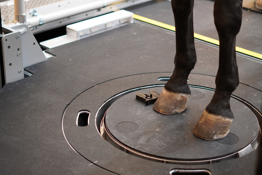
Figure 1: Set-up of a horse for CT imaging of the forelimbs in a natural, weight-bearing position. The CT gantry is housed beneath the system platform and is elevated by the operator to scan the limb pair from proximal to distal.
Why Weight-Bearing CT Matters in Horses
Unlike CT scanners that require a horse to be recumbent under general anesthesia or standing but not bearing weight, the Equina® allows for scanning while the horse remains standing and bearing weight naturally. Figure 1 shows a sedated horse positioned for a standing CT scan. The scanner’s compact design allows imaging up to the mid-radius or mid-tibia, while the horse remains in a natural, weight-bearing stance. This distinction is critical because:
Accurate Anatomical Representation
Bones, joints, and soft tissues align differently when a horse is non-weight-bearing. Weight-bearing CT captures the structures as they function during movement and while bearing load, leading to more accurate diagnoses. (Figure 2)Improved Safety
By avoiding general anesthesia, risks associated with recovery, injury, and post-anesthesia complications are greatly reduced. Additionally, limbs do not have to be placed in an unnatural non weight bearing position through a vertical gantry opening while putting the safety of the horse at risk.Faster Diagnosis
Standing scans can often be performed in a fraction of the time, streamlining clinical workflow and minimizing stress for both horse and handler.

Figure 2: Pre-surgical P2 fracture in QH horse stallion.
Importantly, it makes sense to evaluate certain pathologies such as cartilage and bone damage, or hoof balance in a natural weight bearing position. If both limbs of the horse can be scanned at the same time in weight bearing circumstances like the Equina by Asto CT, that is even better. Both limbs can be compared, and subtle differences can be noted. (Figure 3)

Figure 3: Comparison of left and right carpus under identical conditions. Lesions are shown in both radial carpal and 3rd carpal bones in both midcarpal and carpometacarpal joints.
How Equine Weight-Bearing CT Compares to Human Technology
In human medicine, weight-bearing CT (WBCT) has already revolutionized orthopedic imaging especially in podiatry, sports medicine, and orthopedics. Surgeons use WBCT to assess real-time joint alignment, ligament tension, and bone positioning under physiological load (Rojas et al., 2022; Belvedere et al., 2020).

Figure 4: Comparison of left and right distal tarsus, affected vs. unaffected under identical conditions. The left tarsus shows joint space collapse and osteoarthritis.
Similarly, Equina® brings these same benefits to equine veterinary medicine, allowing veterinarians to evaluate:
Subtle joint space changes in the hoof, pastern, fetlock, hock, and carpus (Figure 4).
Pre- and post-surgical planning to ensure the horse’s natural bone structure, function and movement is restored. (Figure 5).
Biomechanical understanding for performance horses, where even small changes in load distribution can impact athletic potential.
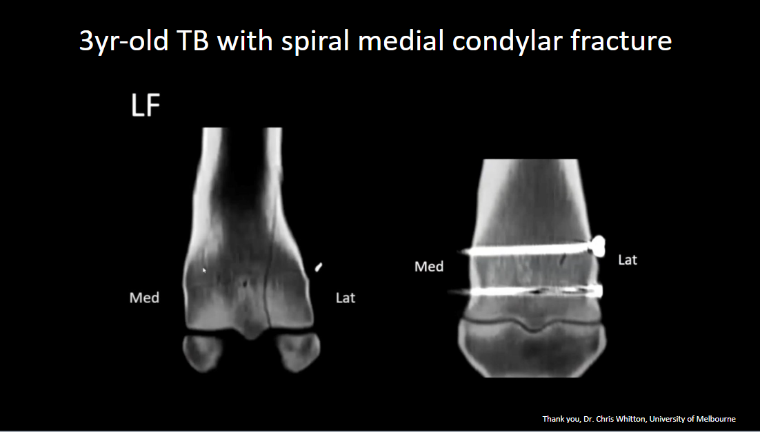
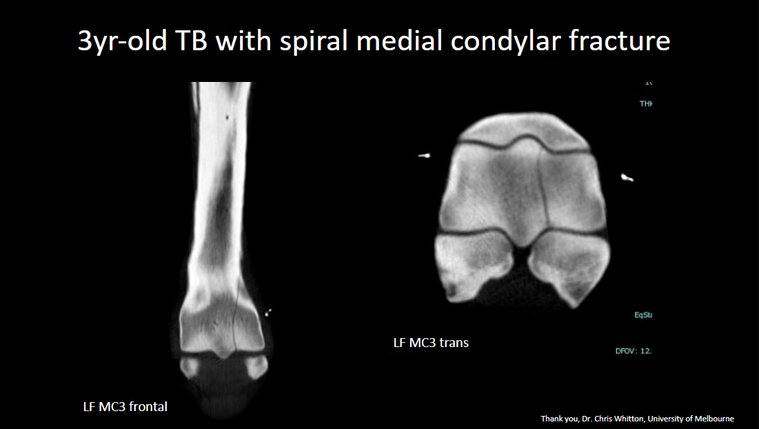
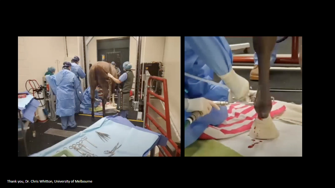
Figure 5: 3-year-old TB with left front spiral medial condylar fracture. CT images show the fracture pre-surgical and screw placement post-surgical.
Where non weightbearing CT might miss dynamic, load-bearing pathologies, the Equina® makes them visible similar to human WBCT for conditions such as early-stage arthritis, hallux valgus, or ankle instability.
Key Benefits of the Equina® Standing CT
Non-Invasive & Low-Risk – No need for general anesthesia; sedation is all that is needed.
High-Resolution Fan-Beam Technology – Delivers superior image clarity compared to standing cone-beam alternatives.
360° True Cross-Sectional Imaging – Provides comprehensive diagnostic detail of the equine distal limb and head.
Improved Case Throughput – Faster scans mean more cases can be diagnosed in less time.
Enhanced Client Communication – Clear, 3D images help owners and referring veterinarians better understand the diagnosis and treatment plan.
Transforming Equine Care—One Scan at a Time
The parallels between human and equine weight-bearing CT are clear: both allow clinicians to view the body under real-world conditions, improving accuracy, reducing risk, and enabling earlier intervention. With the Equina®, veterinarians can bring this standard of care directly to their patients, revolutionizing equine imaging and improving outcomes.
References
Belvedere C, Ensini A, Catani F, Leardini A. Correlations between weight-bearing 3D bone architecture and dynamic plantar pressure measurements in the diabetic foot. J Foot Ankle Res. 2020;13:31. doi:10.1186/s13047-020-00431-x.
Brounts SH, Lund JR, Whitton RC, Ergun DL, Muir P. Use of a novel helical fan beam imaging system for computed tomography of the distal limb in sedated standing horses: 167 cases (2019–2020). J Am Vet Med Assoc. 2022;260(11):1351-60. doi:10.2460/javma.21.10.0439.
Rojas D, Mansur NSB, Dibbern K, Auch C, Schmidt B, Vivtcharenko V, Li S, Phisitkul P, de Cesar Netto C. Weightbearing computed tomography for assessment of foot and ankle deformities: The Iowa experience. Foot Ankle Int. 2022;43(11):1520-31. doi:10.1177/10711007211044806.