ASTO CT / BUILT AROUND THE HORSE
What to Consider When
Purchasing a Standing CT Scanner
Why purpose-built equine imaging matters for the horse, the clinical team, and the practice.
Standing CT is proven. The next decision is choosing a system that was designed around the horse — rather than asking the horse, the staff, and the facility to adapt to the machine.
Purchasing a standing CT scanner is a significant clinical and business decision. Image quality matters, but it is only one part of the evaluation. The right system should also support safe patient positioning, efficient workflows, manageable installation, confident operation, dependable service, and a financial model that fits the practice.
This is where purpose-built design becomes important. Asto CT was created specifically for equine and large animal imaging. Its geometry, scanning platform, power requirements, self-shielding, workflow, and support model were developed around the realities of working with horses. That distinction separates a premium equine CT system from a human scanner that has been adapted for veterinary use.
“It makes sense that any equine practice with an advanced lameness program would have Equine Standing CT installed. For us at Virginia Equine Imaging, it's a critical tool for diagnosing lameness and developing treatment plans. We're seeing more detail, reaching diagnoses faster, and returning horses to their owners with clear answers.”
Dr. Kent Allen — Veterinarian & Owner, Virginia Equine Imaging
Start with the horse,
not the specification sheet
A scanner may produce excellent images in a human hospital and still create practical problems in an equine environment. Horses vary in size, flexibility, temperament, pain level, and ability to tolerate unfamiliar positioning. A clinically useful system must account for that variation from the start.
Some CT systems adapted from human imaging platforms may require positioning approaches based on scanner geometry — imaging one leg at a time, a farrier-like stance, or aligning anatomy to the scanner's field of view. Depending on the patient and clinical presentation, these requirements may influence exam workflow, motion management, scan coverage, and ease of imaging in horses.
Age and lifecycle should also be examined closely. Some human CT platforms reach veterinary practices after a decade or more of prior use. Buyers should ask when the system was manufactured, how long key components have been in service, and which parts remain supported. The system should also be engineered for 24/7 operation, with a strong focus on uptime and serviceability.
| Evaluation area | Question to ask | Why it matters |
|---|---|---|
| Patient positioning | Can the horse stand naturally, or must the horse conform to the scanner? | The system should work around the horse in its natural state of being, not idealized positioning. |
| Anatomical reach | Which limbs, joints, head, and neck regions can be scanned in a standing patient? | Equina scans paired limbs vertically and the head and neck horizontally, with standing neck reach to approximately C7 in many cases. |
| Sedation and anesthesia | How often is general anesthesia required for routine studies? | Standing imaging under mild sedation can reduce anesthesia use for many cases. Anesthesia is still required for lower neck, stifle, and pelvis scans. |
| Patient variability | Can the system accommodate painful, older, large, or temperamental horses? | Purpose-built geometry is intended to reduce forced positioning and motion. |
Evaluate workflow and efficiency
from arrival to report
Workflow is one of the clearest differences between CT systems. A purchase decision should account for the entire exam — patient preparation, positioning, image acquisition, repeat scans, staffing, post-processing, and reporting.
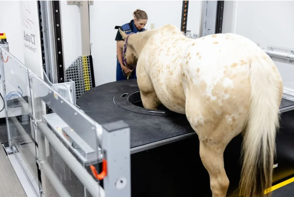
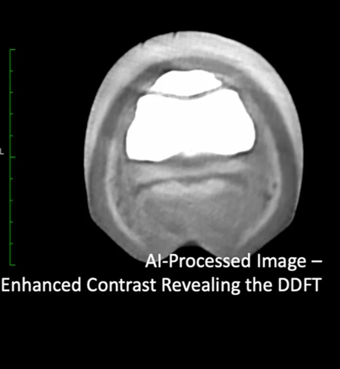
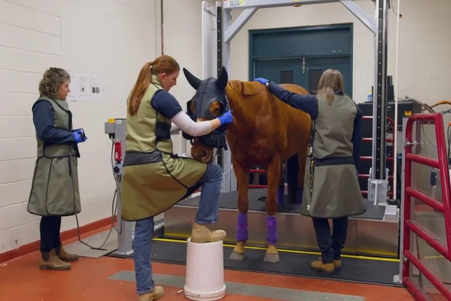
Asto CT is designed for a straightforward weight-bearing workflow. Horses walk onto the platform, two limbs can be scanned at the same time, and most patients can be scanned once without repeat positioning or general anesthesia. AI-driven diagnostic imaging and modern reconstruction tools can further support speed, efficiency, and diagnostic confidence — drag the slider below to see it in action.
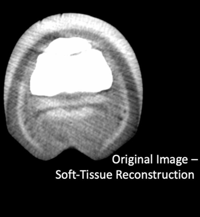
“Our efficiency has soared; what used to take days now takes minutes. As technicians, the CT has empowered us to independently manage scans, enhancing our role in patient care while allowing our veterinarians to focus on more cases.”
Shannon Hullett — Large Animal Surgery Technician, UMN Leatherdale Equine Center
| Workflow stage | What to measure |
|---|---|
| Patient preparation | Sedation plan, loading, positioning, and safety checks |
| Scan acquisition | Time to position, anatomy captured, and frequency of motion-related repeats |
| Staffing | Number of people required and the level of specialist training needed |
| Image handling | Reconstruction speed, AI support, PACS integration, and remote review |
| Throughput | Realistic number of cases per day, not only scanner acquisition time |
| Clinical handoff | How quickly images and reports can be shared with the radiologist or primary veterinarian |
Look beyond purchase price
to installation complexity
The scanner is only one part of the investment. Facility modifications, power, shielding, HVAC, patient access, construction management, and commissioning can materially affect the final cost and timeline.
Human CT systems may require substantial three-phase power and specialized cooling — common in human hospitals but less practical in equine practices. Equina requires a 240V single-phase, 30A power supply and space equivalent to two standard stalls. Installation, testing, and training are typically completed in under two weeks, depending on site readiness.
| Installation category | Questions for the vendor |
|---|---|
| Room and access | Recommended room dimensions, ceiling height, doors, horse entry paths, and unloading route |
| Construction | Pit dimensions, embedded conduits, drainage or sump requirements, and floor finish |
| Electrical | Power phase, amperage, auxiliary outlets, backup power, and upgrade cost |
| Radiation | Shielding assessment based on wall construction and expected use |
| Environmental durability | Protection from dust, liquids, horse kicks, and daily barn conditions |
| Project support | Project coordinator, site visits, checklists, timeline ownership, and issue escalation |
Assess ease of use,
training, and long-term support
A premium scanner should be a dependable, practice-wide tool, not a system limited to a single operator. Buyers should evaluate training, onboarding, ongoing support, and how the vendor helps the team improve protocols over time.
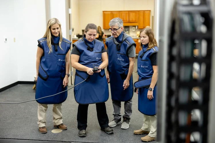
Asto CT provides hands-on training for veterinarians, technicians, practice managers, and other staff. Ongoing applications support helps refine imaging protocols and clinical workflows, while remote and on-site service minimizes downtime and maintains facility control over system access.
The vendor relationship should also include preventive maintenance, software updates, parts availability, and a clear response process — particularly important when comparing a current purpose-built platform with a repurposed system whose original manufacturer may no longer prioritize an older model.
Match the system
to the practice model
No two practices are set up the same; imaging centers, smaller practices, and referral hospitals each have different business objectives when it comes to justifying the cost and benefit of CT. Practices should consider the revenue models that best align with their goals — serving as a regional imaging center, participating in collaborative referral networks, or maximizing retained case revenue through ancillary services.
Best fit: An imaging service that returns the case to the primary veterinarian.
Referred cases are imaged and returned with images and a report — supporting clear professional boundaries and CT revenue without competing for additional care. This positions the practice as a leading regional imaging center.
Clarify: reporting turnaround, communication boundaries, scheduling, and case transfer process.
Best fit: Regional partnerships with complementary diagnostic or treatment services.
Smaller practices can expand access to advanced diagnostics by partnering with nearby practices offering complementary imaging such as PET, bone scan, and standing MRI — keeping each practice focused on its strengths.
Clarify: shared protocols, transport, report access, and clear ownership of follow-up care.
Best fit: Practices maximizing the clinical and financial value of advanced diagnostics through expanded treatment offerings.
One of the most overlooked benefits of a standing CT scanner is its ability to increase case retention and generate downstream clinical opportunities. When veterinarians have greater diagnostic confidence, they are more likely to proceed with advanced treatment rather than pursue extended conservative management or refer the patient elsewhere.
Standing CT functions as a diagnostic gateway — identifying the right treatment path and keeping more of a patient's care (surgery, lameness work, dentistry, sports medicine, regenerative therapy, rehab) inside the practice.
Clarify: historical case retention rates, surgery conversion rates, capacity, and referral treatment agreements.
| Financial model | Best fit | Items to clarify |
|---|---|---|
| Capital purchase | Established caseload, available capital, focus on long-term ownership and margin | Total cost of ownership, service after warranty, depreciation, and upgrade path |
| Lease | Preserving capital, managing adoption risk, or preferring predictable monthly expense | Terms including service and software, purchase options, and absence of revenue sharing. Equina is non-revenue sharing. |
Use a whole-system
purchasing checklist
A CT purchase should be judged as a complete operating system rather than a scanner alone. The strongest option is the one that improves diagnostic confidence while fitting the horse, the team, the facility, and the economics of the practice.
Confirm before signing
0 / 6 confirmed“Partnering with a vendor like Asto CT was a key aspect to our success throughout the entire process. They provided 360-degree support for every stage, from financing to installation, and the marketing of our scanner.”
Dr. Douglas Langer — Veterinarian (retired), Wisconsin Equine Clinic and Hospital
Built around the horse
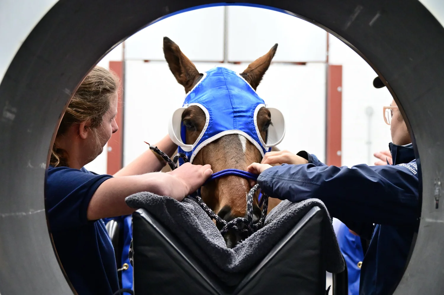
Standing CT has already demonstrated its value in equine practice. The next step is choosing the right execution of that technology. A premium equine CT scanner should do more than create high-quality images — it should make imaging safer, easier, faster, and more sustainable for the practice.
Asto CT was built specifically for horses, from the bi-axial scanning geometry and weight-bearing workflow to the low-power infrastructure, training, and long-term support — designed to improve how the practice images, how the team works, and how the business performs, without forcing compromises on the horse and practice.