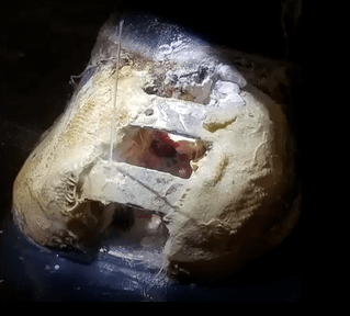
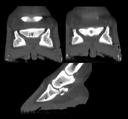
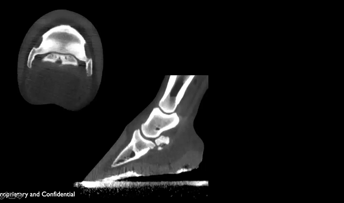
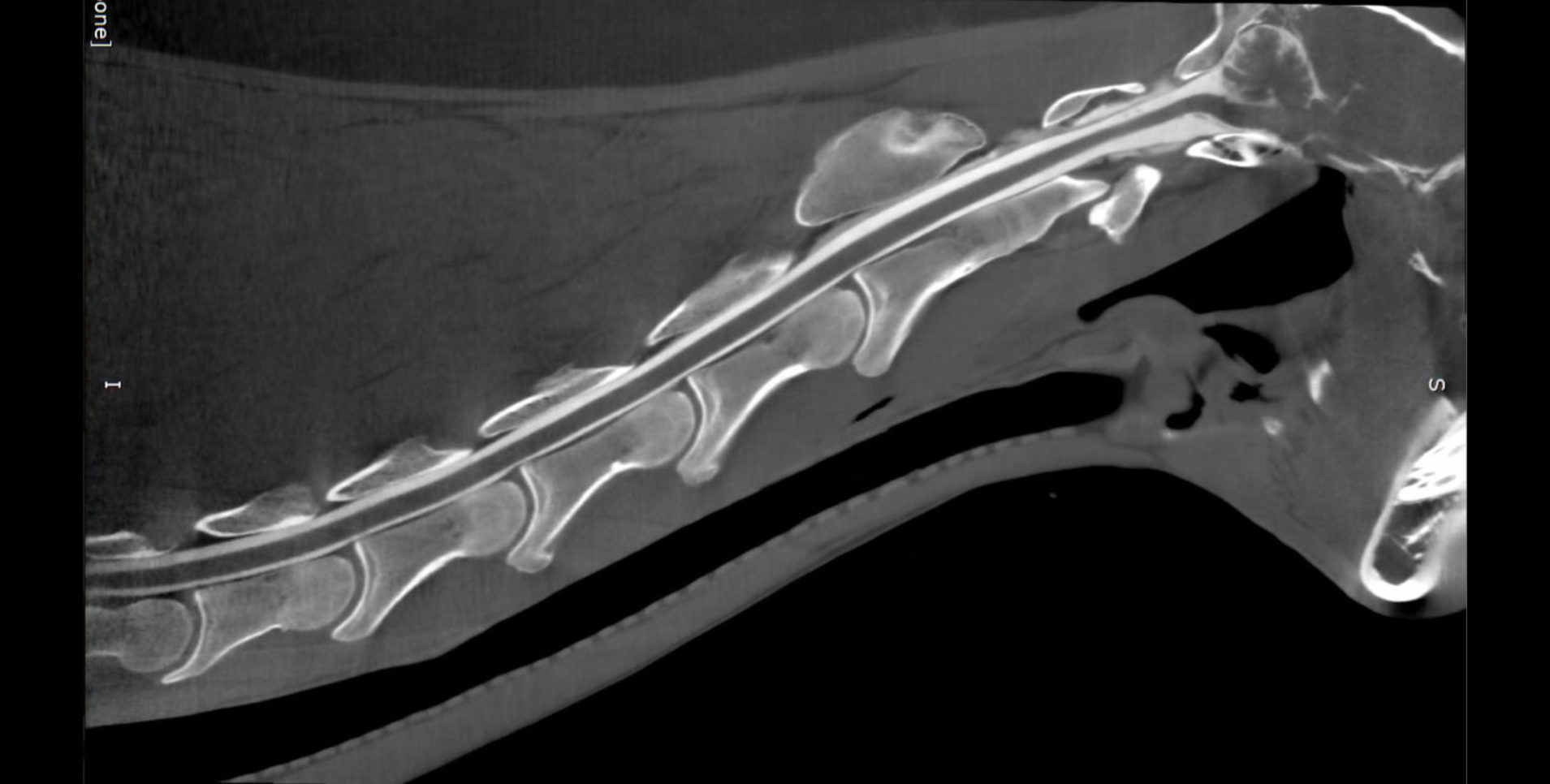
Oliver the 10 yo Tennessee Walker was diagnosed with Squamous Cell Carcinoma causing his left eye to be removed. He was doing well until he went blind in his other eye, CT images show a large mass which was caused from the initial squamous cell carcinoma. Featured: JR Lund, University of Wisconsin-Madison
Read MoreThis 13 yo Shire had a quittor (infected cartilage) which was treated surgically. He then came back a few months later with a persistent draining tract. Featured: Dr. Diego De Gasperi, University of Wisconsin-Madison
Read More13 yo Quarter Horse with a front limb lameness, grade 3 of 5 lameness on the left front, grade 2 of 5 on the right front. Confirmed to be Navicular.
Featured: Dr. Diego De Gasperi, University of Wisconsin-Madison
Read MoreThis is an 11 yo Quarter Horse with a history of chronic lameness in the right front limb, grade 4 of 5 lameness. CT images shows extensive mineralization and confirmed navicular.
Read MoreThis is a 12 yo Thoroughbred that had a right front limb lameness and chronic abscessing. A boney defect was identified in CT images and later confirmed to be a keratoma.
Featured: Dr. Diego De Gasperi, University of Wisconsin-Madison
Read MoreThis is a 7 yo QH mare with a history of forelimb lameness, more severe in the left forelimb. CT images show a osteo-cyst like lesion in the front left navicular bone that is irregular in shape. Featured: Dr. Diego De Gasperi, University of Wisconsin-Madison
Read MoreWe're looking at an 8yo Saddlebred with navicular syndrome that had on and off lameness in the left front forelimb. A palmar digital nerve block was performed to localize the lameness. CT images show large flexor cortical erosion that communicates with the synovial invagination shown in the video. Other CT images show a large cortex defect with surrounding sclerosis and a deep penetrating synovial invagination. Featured: Dr. Diego De Gasperi