5 yr old Quarter Horse with a front limb lameness associated with a 3 cm laceration.
Read More
5 yr old Quarter Horse with a front limb lameness associated with a 3 cm laceration.
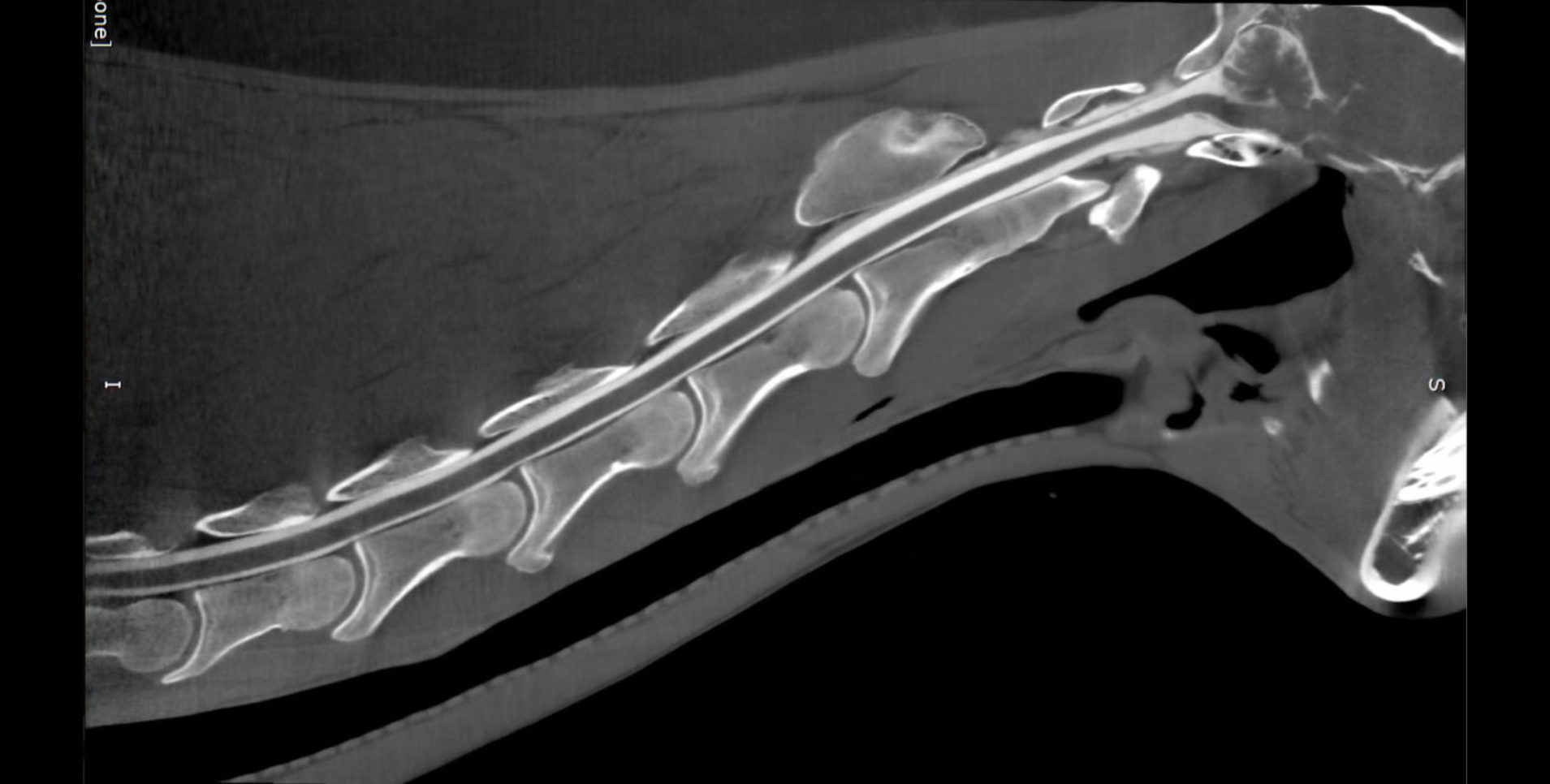
Read MoreIn this video the Equina imaged a tendon lesion, squamous cell carcinoma, and neurologic disorder in the neck.
Read MoreMark is nine-year-old Clydesdale gelding who had a long history of chronic abscessation and white line disease on the left front foot. Mark had several hoof wall debridement's and resections and is currently doing well.
Read MoreThis horse had a history of hock arthritis and lameness. CT images show several cyst-like lesions in the third tarsal bone and the proximal aspect of the third metatarsal bone. The arthritis was noticed mostly in the right hind but also in the left hind with narrowing in the joint space. Featured: Dr. Diego De Gasperi, University of Wisconsin-Madison
Read MoreThese cases show lesions in the fetlock joint area. Case 1 shows lesions with lysis and surrounding sclerosis with hyperattenuating lesions into P1. Case 2 shows a lesion of lysis with surrounding sclerosis in the fetlock joint. Case 3 shows lameness in the right hind fetlock. CT images show subchondral cyst like lesions with lysis and surrounding sclerosis. Featured: Dr. Diego De Gasperi, University of Wisconsin-Madison
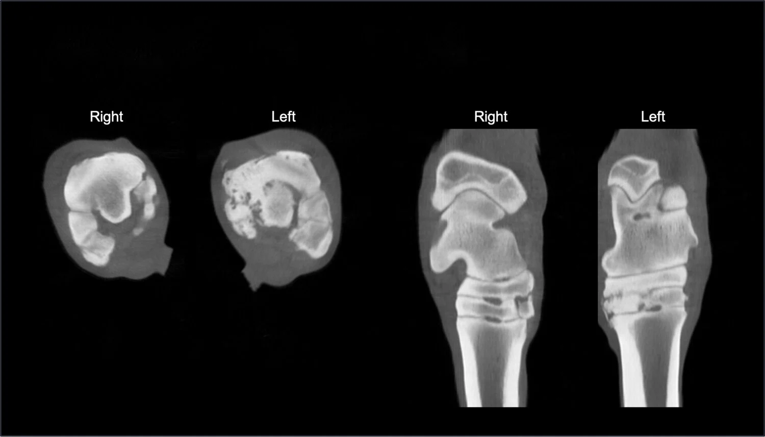
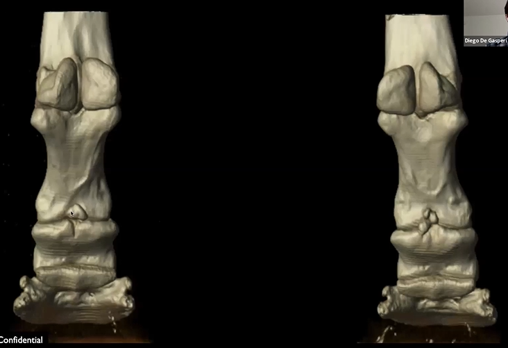
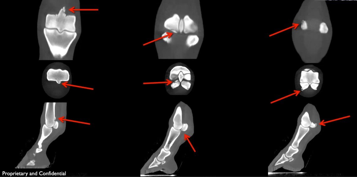
Read MoreCT images show bilateral fragmentation of the proximal palmar articular surface of the middle phalanx. They surgical removed multiple fragments in the right forelimb. Watch the video to view 3D reconstructions. Featured: Dr. Diego De Gasperi, University of Wisconsin-Madison
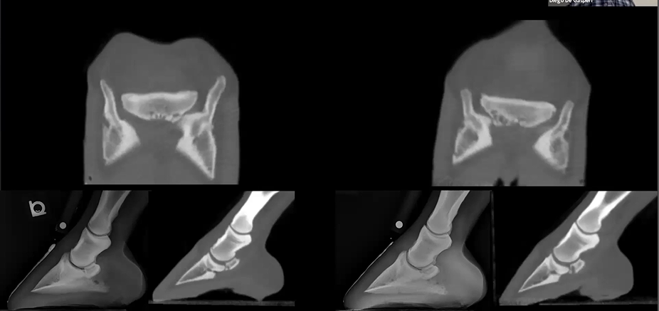
Read MoreThis is a navicular case with more severity in the right front limb. CT images show deep synovial invagination, sclerosis, and distal boarder fragmentation. Featured: Dr. Diego De Gasperi, University of Wisconsin-Madison
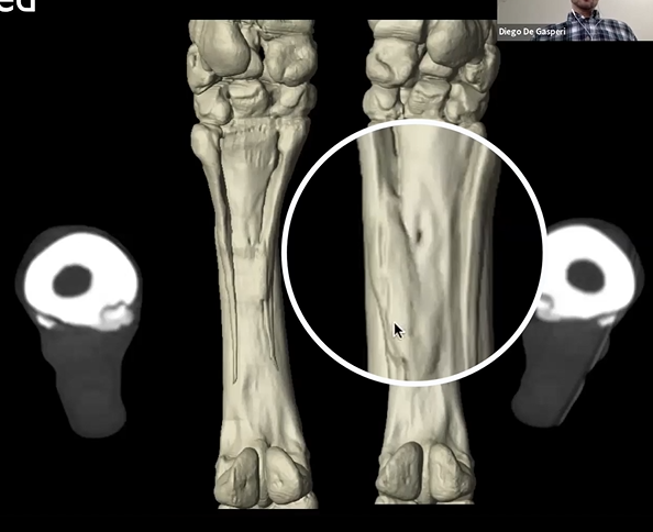
Read MoreThis horse came in for a suspected splint bone fracture. CT images show new bone formation involving the axial aspect of both medial splint bones. Featured: Dr. Diego De Gasperi, University of Wisconsin-Madison
Read MoreThis horse became lame after being lunged in the round pen. The local Vet performed a PD block upon which the horse became sound. CT images show an incomplete fracture of the third phalanx.
Featured: Dr. Diego De Gasperi, University of Wisconsin-Madison
This horse became non-weight bearing on the right hind. Radiographs were taken and the horse was diagnosed with a P3 fracture. CT images show and abaxial articular P3 fracture and a lesion on the coffin bone.
Featured: Dr. Diego De Gasperi, University of Wisconsin-Madison
Read MoreThis horse came in with a open complete comminuted fracture of the lateral splint bone in the right hind limb. A partial ostectomy was performed, removing the bone just above the fracture site. After surgery, discharge was noticed in several wound bandages. CT images show bone sequestrum fragment was found on the axial aspect of the splint bone. Once the fragment was removed the horse healed up nicely. The sequestrum found in CT images was not able to be seen in radiographs. Featured: Dr. Diego De Gasperi, University of Wisconsin-Madison
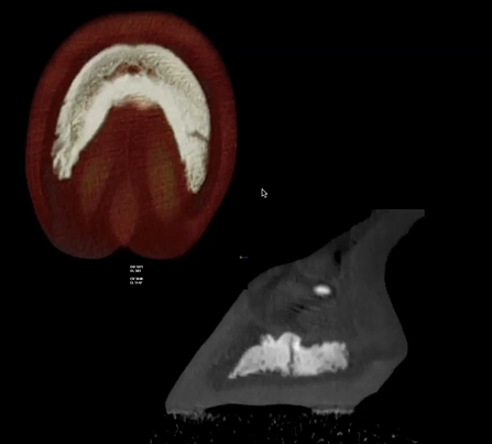
This is a 9 yo hunter jumper that had the 10 tooth extracted and developed an oral sinus fistula. A plug was inserted, meanwhile CT images were taken, and a tract was shown running into her sinus. A protemp bridge was inserted (prosthetic tooth), the bone filled in and healed well. Featured: Dr. Travis Henry, University of Wisconsin-Madison
Read MoreThis horse had a large dental mass located close to the brain stem, in the calvarium, and in the brain. They elected to not do surgery on this horse due to the complexity of the dentigerous cyst.
Featured: Dr. Travis Henry, University of Wisconsin-Madison
Read MoreThis was a younger horse that came with what they believed to be a tooth root infection. Upon CT imaging sequestrum was discovered along the facial crest, and the horse was diagnosed with a skull fracture.
Featured: Dr. Travis Henry, University of Wisconsin-Madison
This was an older mare that sustained trauma to her mandible in the pasture. CT images show a draining tract and sequestrum, the tooth above the draining track was removed.
Featured: Dr. Travis Henry, University of Wisconsin-Madison
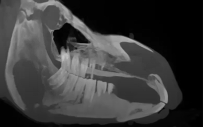
Read MoreThis horse had advanced TMJ pathology, which caused mastication issues. CT images show compression on the temporal hyoid bones and guttural pouch.
Featured: Dr. Travis Henry, University of Wisconsin-Madison
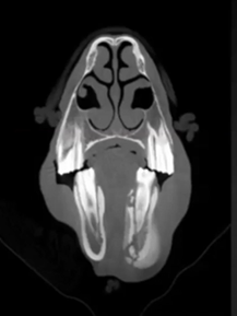
Read MoreThis Warmblood mare came in with sinusitis. CT images show a Diastema which was the cause of the sinusitis. The 10 tooth was removed and Protemp (human crown) was applied to create a prosthetic tooth.
Featured: Dr. Travis Henry, University of Wisconsin-Madison
Read MoreThis horse has all the clinical symptoms of periodontal disease. CT images show an increase in periodontal ligament width, horizontal/vertical bone loss, periodontal infection, diastema, food impaction causing infection to spread and creation of a draining tract.
Featured: Travis Henry, University of Wisconsin-Madison
Read MoreThis middle aged QH presented nasal discharge, believed to be sinusitis. Upon CT scans dense dental material was found in the nasal passage. This material was causing the nasal discharge.
Featured: Dr. Travis Henry, University of Wisconsin-Madison
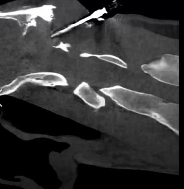
Read MoreThis 18 yo Arabian gelding had a record of cranial nuchal bursitis and a persistent draining fistula. CT images show the osteomyelitis of the occipital bone (severe lysis).
Featured: Dr. JR Lund, University of Wisconsin-Madison