The Equina® by Asto CT
WE COVER THE HORSE FROM HEAD TO HOOF WITH TRUE WEIGHT-BEARING, FAN-BEAM CT

WE COVER THE HORSE FROM HEAD TO HOOF WITH TRUE WEIGHT-BEARING, FAN-BEAM CT
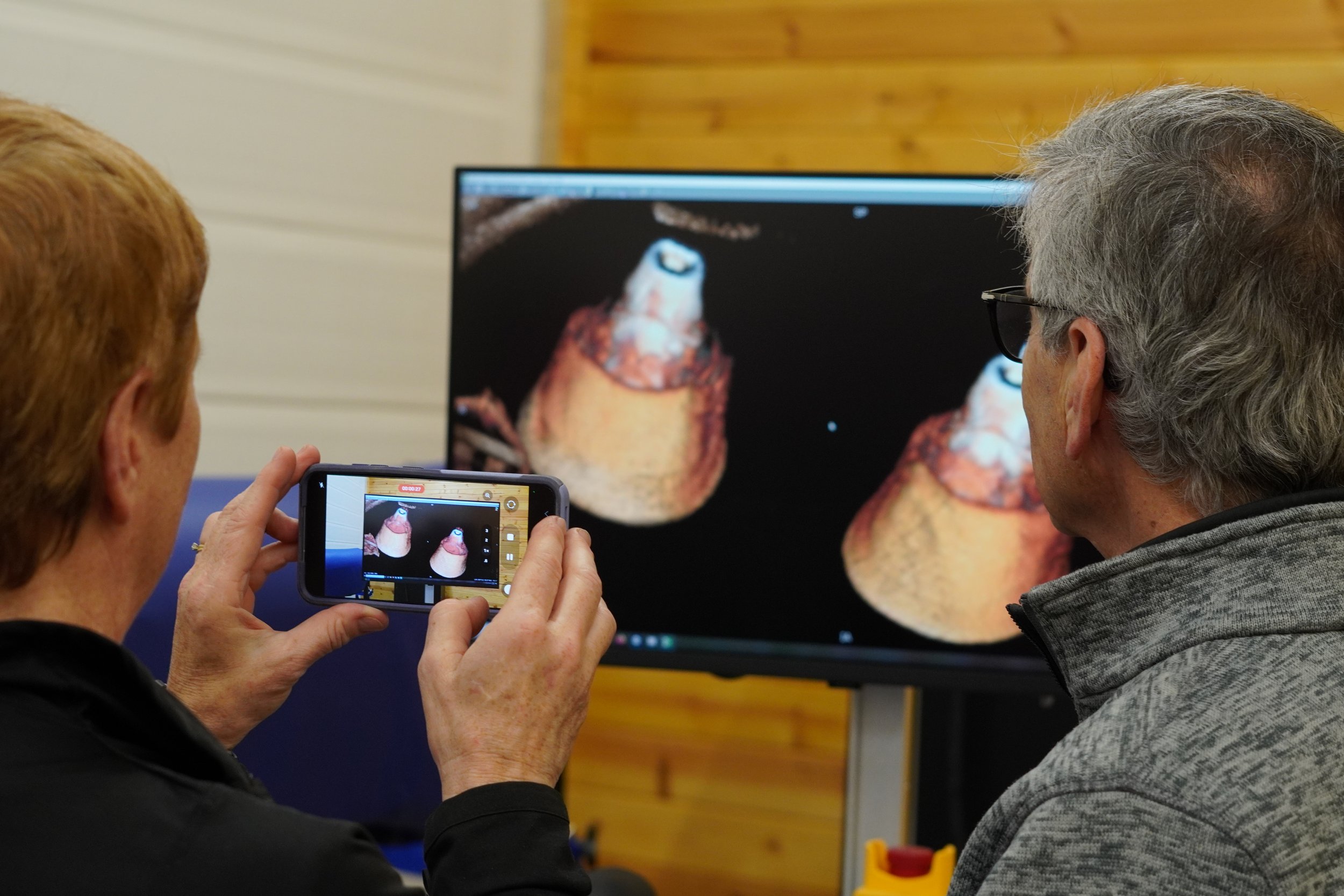
Designed for the equine athlete, Asto CT delivers precise, standing CT scans that support faster diagnosis and better-informed treatment decisions—empowering veterinarians with the clarity they need, when they need it most.
A team of medical imaging experts with unrivalled experience and a strong track-record supporting you at every step of your journey.
Options to purchase the CT scanner outright vs. subscription to meet your clinic’s unique needs.
People you can count on to deliver best-in-class service to your CT scanner with fast respond times.
A community of CT users to share learnings and help you advance your practice.
An unwavering commitment to your project success.
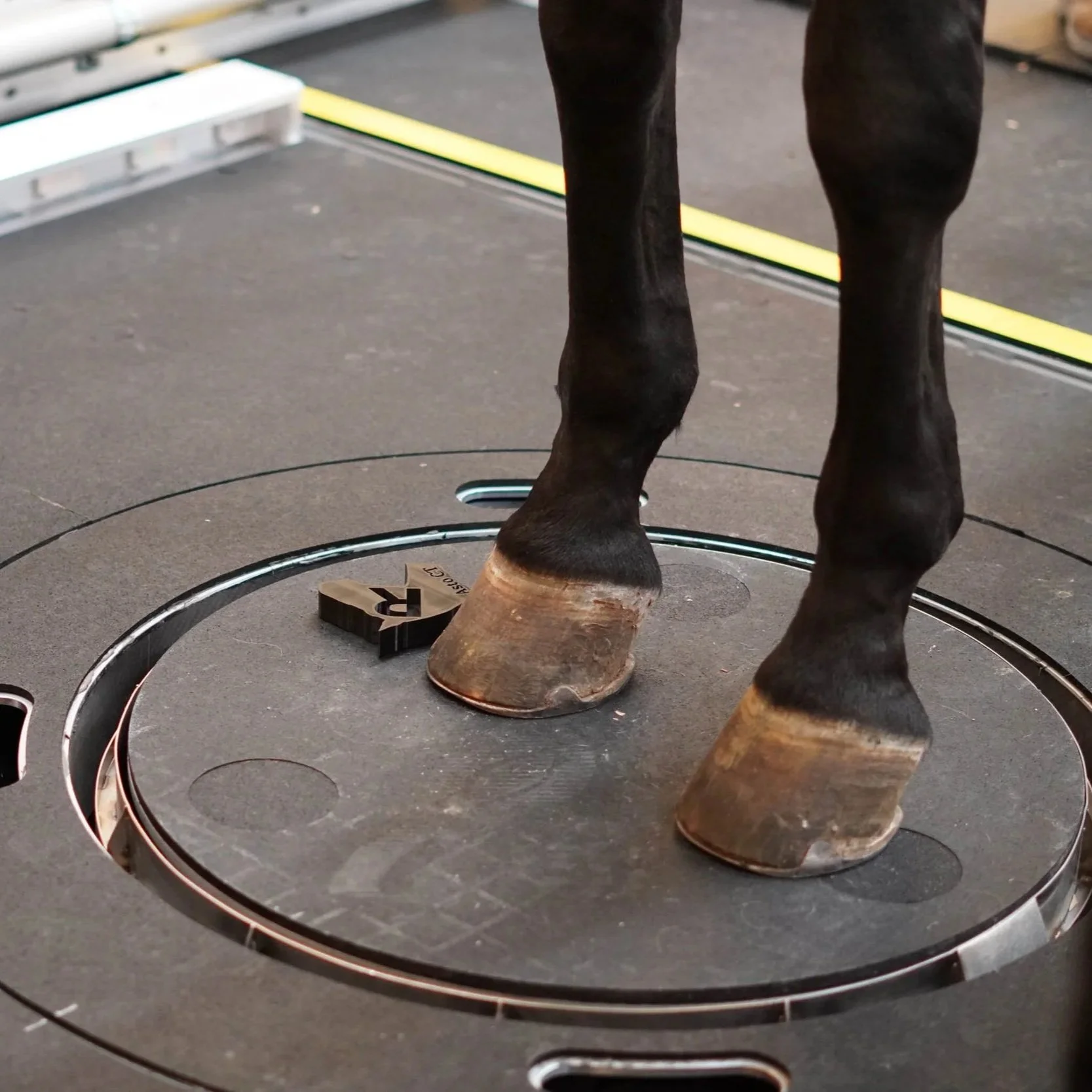
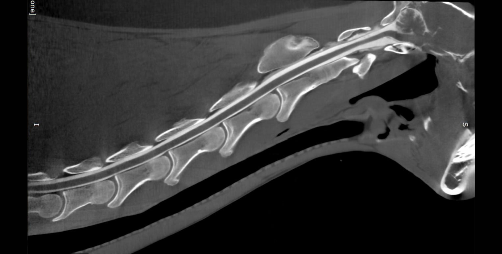
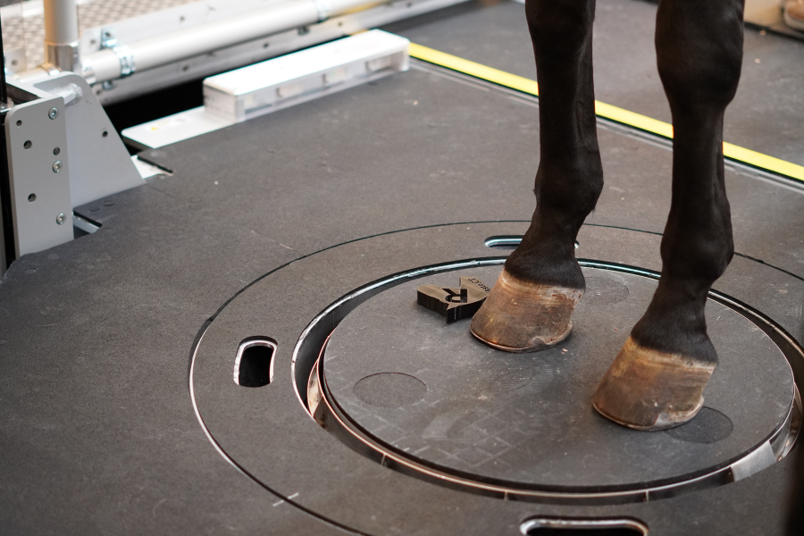
The Equina by Asto CT is the world’s only bi-axial equine standing CT scanner that scans vertically for limb pairs and horizontally for the head and neck. The ability to scan a standing sedated horse eliminates the need for general anesthesia.
CLICK THE ICONS BELOW


Subscribe to our Newsletter – Get the latest updates and exclusive offers delivered straight to your inbox.