

Helping Clinicians Save Time and Hassle with the Convenience of the Equina®


ENHANCING YOUR CLINIC WITH THE EQUINA® STANDING CT AND THE PATH TO IMPLEMENTATION
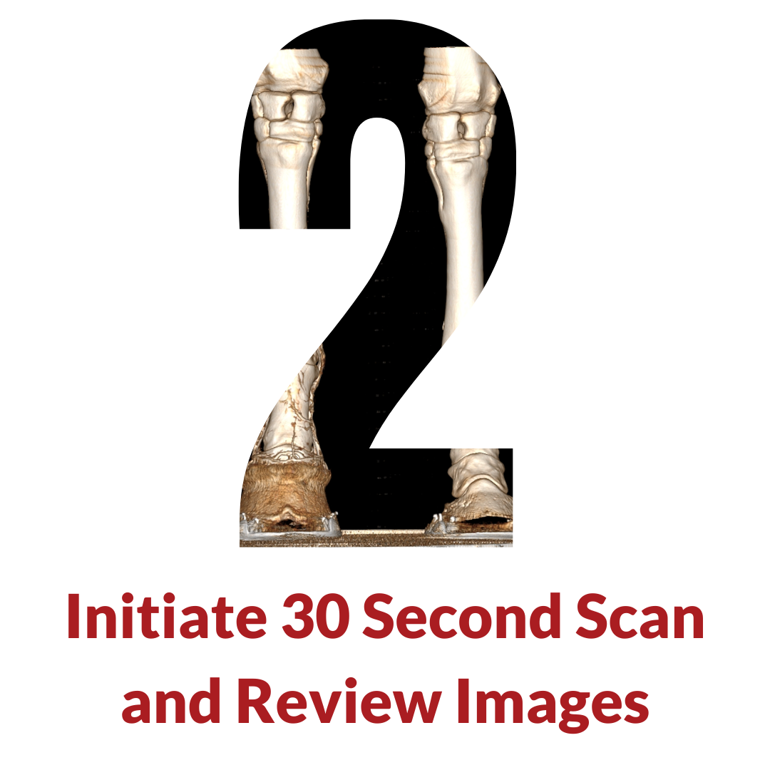
The successful integration of equine standing CT into veterinary practice requires careful planning and preparation. Here are key steps to consider when implementing this technology.

WHITEPAPER: INJURY PREVENTION IN THOROUGHBRED RACEHORSES USING SAFE, HIGH-QUALITY STANDING CT IMAGING
The use of this advanced veterinary technology for early detection and management of musculoskeletal injuries can move us towards a new standard of care for Thoroughbred racehorses. In this paper, we provide a brief description of the key features of the Equina® system, and its use in pre-race screening.

WHITEPAPER: A NOVEL HIGH-QUALITY EQUINE STANDING CT SCANNER: THE ASTO CT EQUINA®
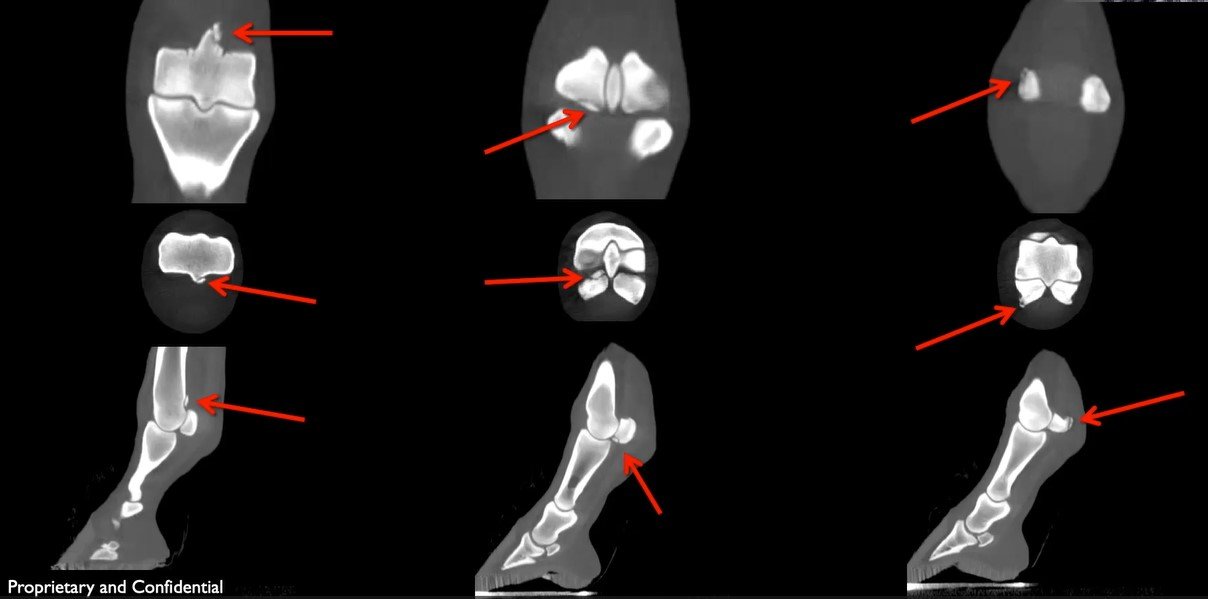
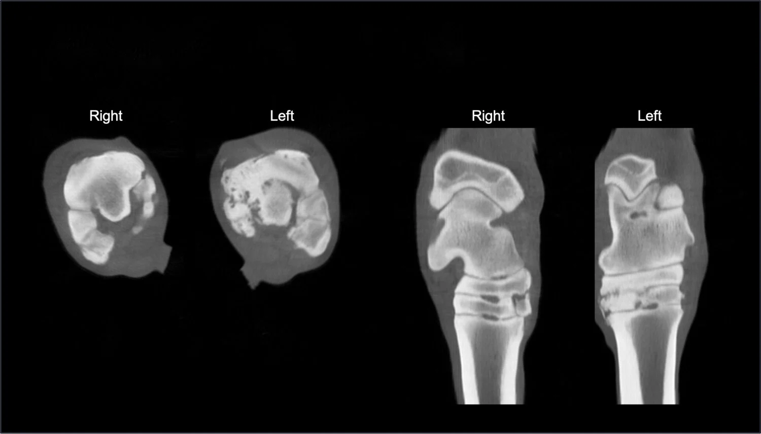
"When a comparison was made between cases that had both radiographs and a CT scan performed, only in 25% of these cases (22/88) was the diagnosis made with radiographs, indicating up to a 75% false negative rate for radiographs alone."

WHITEPAPER: FAN BEAM CT VERSUS CONE BEAM CT FOR VETERINARY MEDICINE
Fan-beam systems offer faster scan times which make its images less susceptible to motion artifacts due to swaying or other subtle movements of the equine patient. Fan-beam images also have higher image quality, enabling them to be used to identify subtle pathological changes in the limbs of horses, and important advantage over both traditional radiography and cone-beam CT scanning.
featured case studies


EQUINA® IN CLINICAL PRACTICE
Since November 2018, the Equina® sites have completed more than 5,000 scans on diverse patients, including clinical cases. With proven sedation protocols and the simplicity of using Equina®, patients are in and out in 10 minutes.


Current Asto CT Sites
Frequently Asked Questions
-
The Equina® by Asto CT is a state-of-the-art robotically positioned computed tomography (CT) scanner for imaging a sedated, standing horse – no anesthesia is required.
-
The Equina® is able to quickly & safely evaluate:
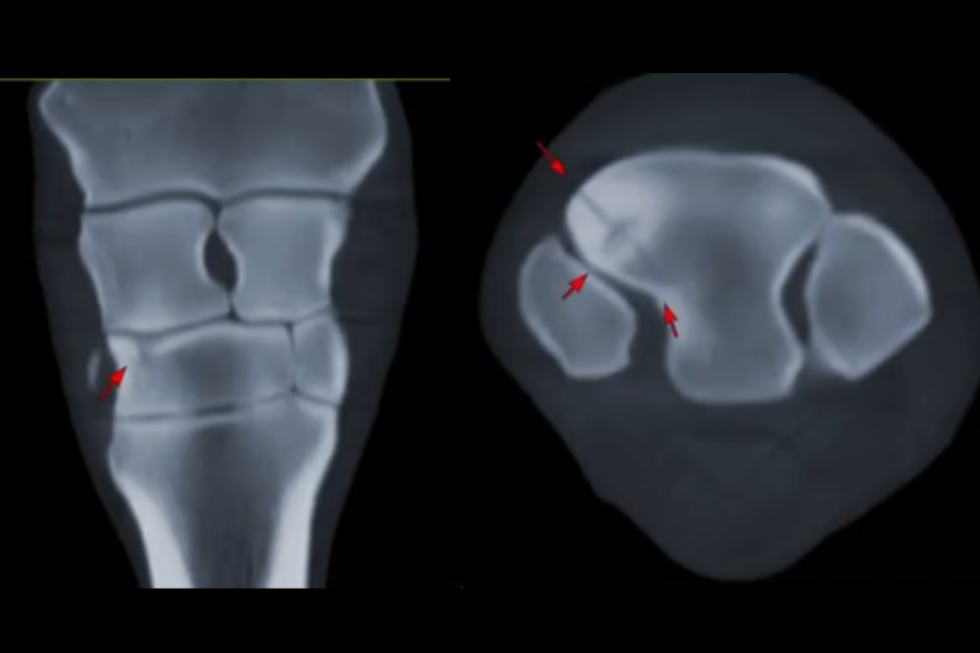
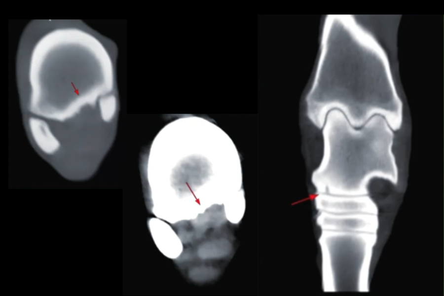
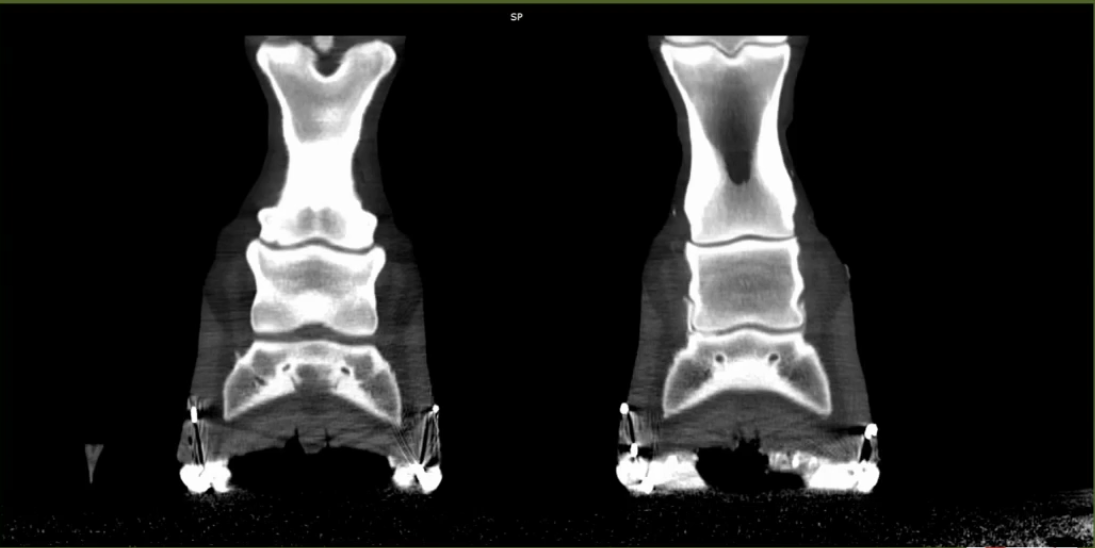
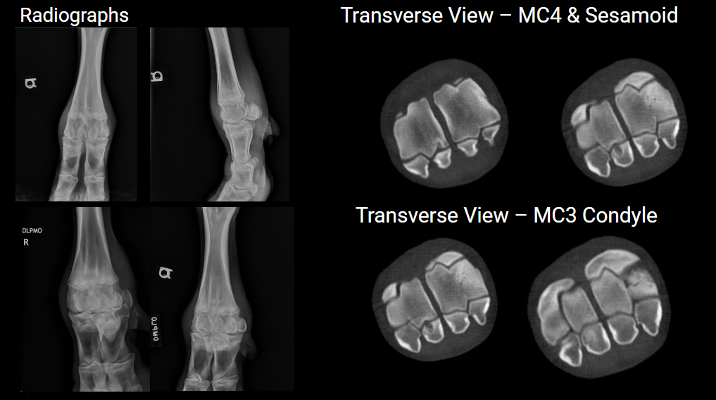
Proximal Limbs: Limb pairs can be imaged from carpus distally to the foot in the thoracic limb, and from tarsus distally to the foot in the pelvic limb in a scan time of 20-30 seconds. Indications for use include: Pre-purchase screening of horses; screening of horses during athletic events, such as jumping & eventing; and diagnosis of lameness problems in thoracic or pelvic limbs.
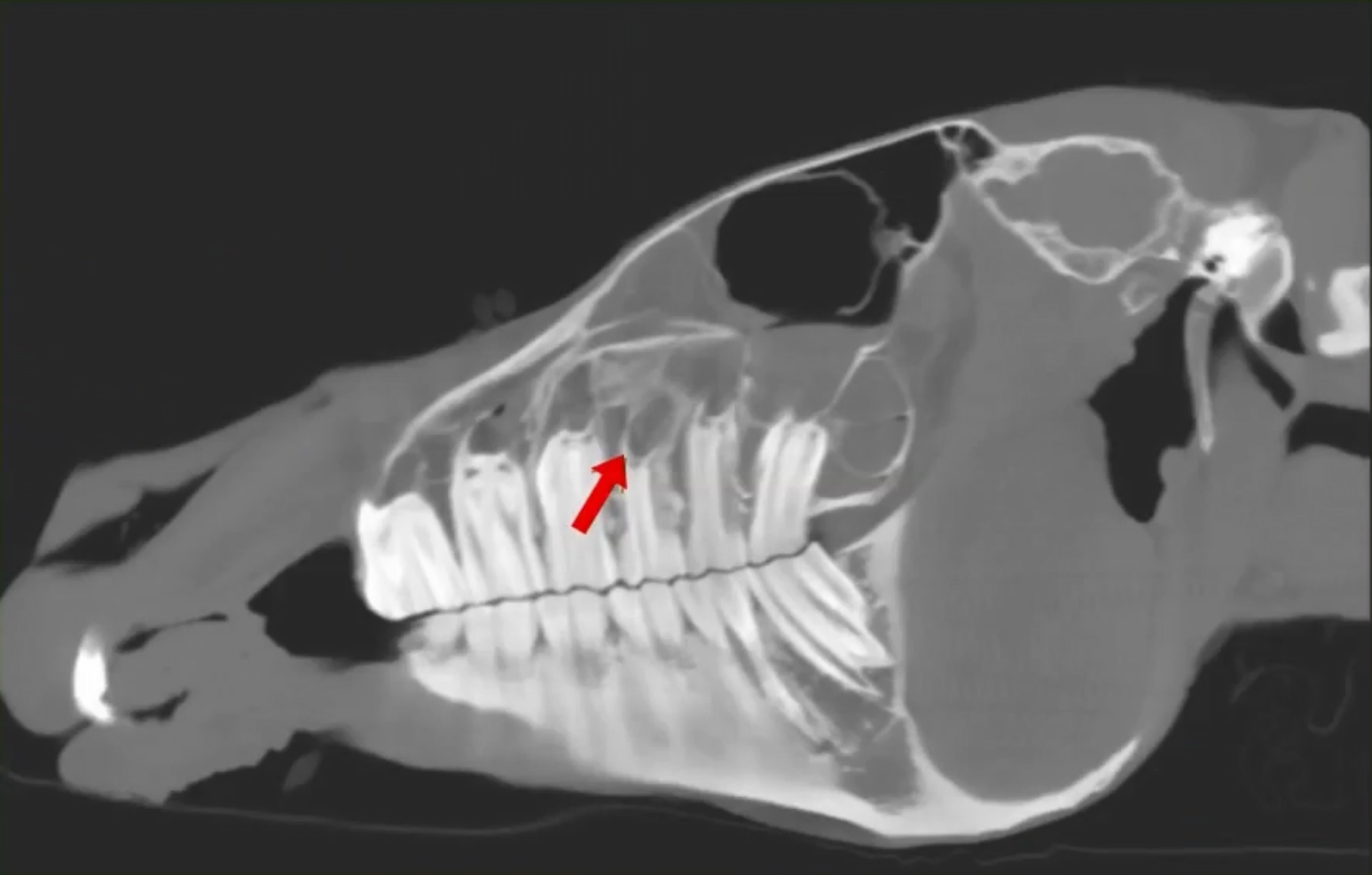
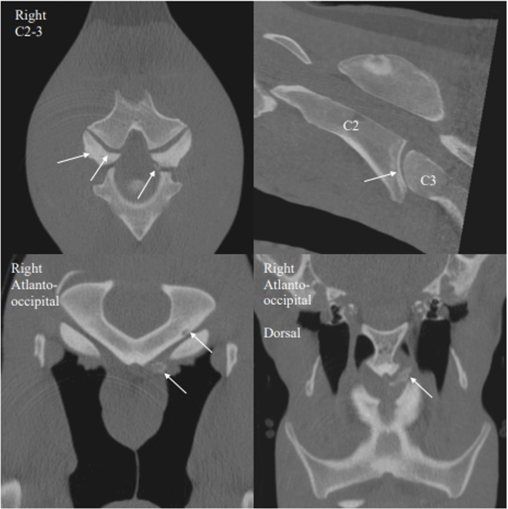
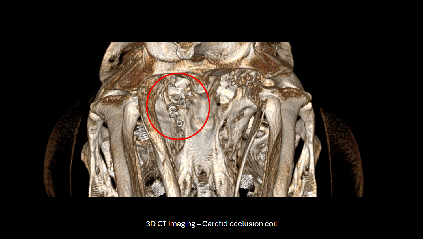
Head & Neck: The head & neck can be imaged from the neck rostrally to the nose with a scan time of 30-40 seconds. Indications for use include: Diagnosis of equine diseases of the head, such as ethmoid hematoma, sinus problems and teeth/jaw problems; and diagnosis of equine neck diseases, such as osteoarthritis and fractures.
Imaging under General Anesthesia: Large or small animals can be imaged under general anesthesia on a moveable transport/operating table in lateral or dorsal recumbency. CT scanning of an equine patient under general anesthesia may be useful for preoperative planning immediately before surgery or for special procedures, such as myelography.
-
The Equina has been purpose-built in consultation with equine veterinarians to rapidly acquire CT images from equine patients of all conformations & sizes. The overall strength of the system includes consideration for the weight & power of the horse even in the extreme conditions of kicking or rearing up. Horse-oriented design has informed the choice of colors, materials, sounds, and environmental conditions compatible with horses. The Asto CT Equina is NOT a repurposed human-use CT scanner.
-
The Equina starts with all of the modern features found on the most advanced fan-beam CT systems: helical scanning; multi-row detector configuration (24 rows); high-speed processing (36 slices/rotation); sub-millimeter resolution. Unlike human scanners, Equina’s patented CT architecture allows for the patients to stand anywhere within the 75cm field of view. This eliminates the need for scout scans, reducing dose and procedure time.
-
Flexible: Equina is designed to enable the two most common clinical use cases: standing limb pairs and head & neck scans with the flick of a switch making it a cost-effective solution for most full-service equine hospitals & clinics.
Diagnostic: Equina has best-in-class Standing CT acquisition along with equine-specific image processing algorithms for bone & soft tissue.
Simple: All motions & scanning are initiated from a push button controller. This allows the operator to focus 100% of his/her attention on the patient & handlers in the room for maximum safety.
Rugged: Equina has been shielded and sealed to ensure that it can withstand the equine environment, including unforeseen events like rearing and kicking.
Safe: The Equina runs at very low x-ray dose levels allowing handlers to safely stay in the room with the patient while scans are being conducted.
-
Due to the rapid scan time, movement is not usually an issue. If movement is detected, the operator can move the CT gantry back to the start position and start again. In the rare case of large movements, Equina has a special abort function to stop the scan immediately and rapidly lower the CT gantry.
-
The CT can provide scans of both limbs at the same time, regardless of if the horse is weight bearing or not. That gives you the information to compare both limbs [affected and unaffected] to find a cause for issues. The horse stands exactly like it normally does … If you had to scan one limb at a time, you would never be able to create the exact circumstances for the limbs.
-
Because Equina can generate diagnostic results in only minutes it reduces the need for time-consuming x-ray imaging. This both increases throughput for traditional candidates for diagnostic imaging and increases the number of horses who may be candidates for diagnostic imaging by providing a safe, simple option.
-
The breakeven depends on a lot of different factors, but in general, sites are seeing a breakeven of 12 patients per month with an average client fee of $1000.
-
Once the equipment is on site, it typically takes 2-3 days to install, 1-2 days to commission, and one week to train – the installation can be completed in just 2 weeks.
-
Because space in equine facilities is tight, the Equina has been designed to fit in a compact space. It will be necessary to construct a 32-inch pit with machinery base so the scanner can sit below the limbs of the standing horse. Asto CT can generate facility plans and will work closely with facility personnel to make sure the room remodeling is done correctly.
-
Asto CT offers a comprehensive service plan for maintaining top-level performance, including scheduled maintenance, software upgrades (new features and performance enhancements), and remote support (both telephone and remote login for diagnostics & repairs). Internet access is required for remote login.
-
All Equina purchases include initial training at the time of installation. Asto CT’s remote support allows Asto CT to provide additional training for any questions that may arise and for new features that come with software upgrades.
Get started

Let’s Talk
Let’s discuss if the Equina is right for your clinic, we're committed to helping you make an informed decision. Don't hesitate to contact us so that we can assist you in this process.

Plan Your Installation
We can generate facility plans and will work closely with facility personnel to make sure the room is remodeled for optimal usage.

Install The Equina
With our team of experts, installation takes only 2-3 days, 1-2 days to commission, and one week to train – the installation can be completed in just 2 weeks.
