Dr. Kevin Keegan
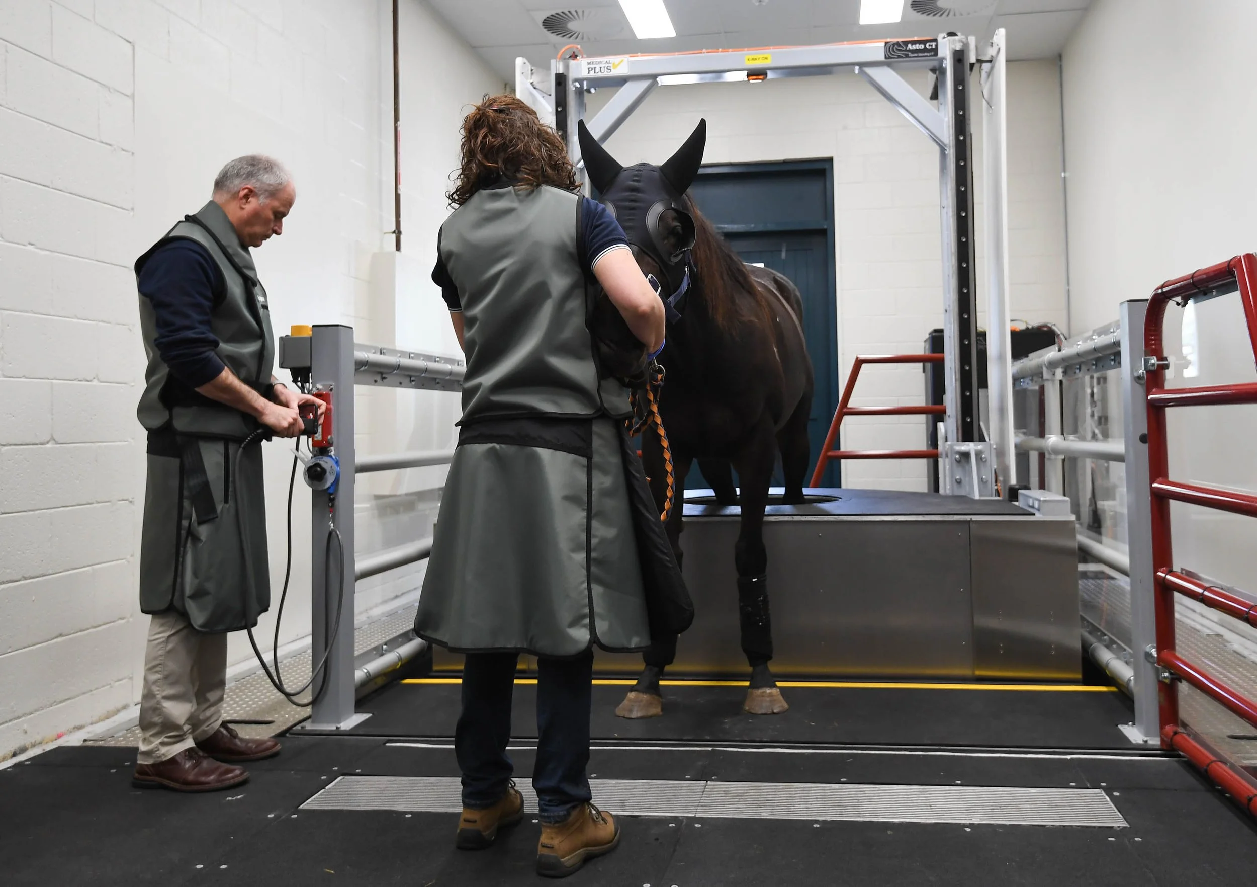
“Getting a study done with the Asto CT is incredibly easy. Beyond the superior image quality and expanded diagnostic capabilities compared to standard X-rays, it’s a true labor-saving device”
Read more →
Dr. Kevin Keegan
DVM, MS, DACVS Professor, Equine Surgeon, MU VHC Equine Hospital
Read More
Dr. Kent Allen
“It makes sense that any equine practice with an advanced lameness program would have Equine Standing CT installed. For us at Virginia Equine Imaging, it’s a critical tool for diagnosing lameness and developing treatment plans. ”
Read more →
Dr. Kent Allen
DVM, CERT. ISELP, President & Executive Director ISELP, Virginia Equine Imaging
Read More
Dr. Sabrina Brounts
“Traditional CT does not allow for the close observation that the Equina provides. With traditional CT there is increased risk, if you’re leaving a horse tied to a machine, or positioned in an unnatural position”
Read more →
Dr. Sabrina Brounts
DVM, MS, DACVS, DACVSMR (Equine Clinical Professor, Head of Large Animal Surgery, UW Madison)
Read More
Dr. Chris Whitton
“Imaging both limbs simultaneously helps us identify unexpected lesions with significant prognostic implications. It allows for a complete assessment of key joints, and provides a view of bone, soft tissues, and foot balance.”
Read more →
Dr. Chris Whitton
BVSc, FANZCVS, PhD, Professor at Melbourne Veterinary School
Read More
Dr. Nicolas Ernst
“The biggest benefit of the standing CT at our clinic is its speed and precision. Compared to other imaging modalities like MRI and X-ray, we can obtain high-quality, three-dimensional diagnostic images in under a minute”
Read more →
Dr. Nicolas Ernst
DVM, DACVS-LA, Associate Professor VPM, Equine Surgeon at the UMN Leatherdale Equine Center
Read More
Megan J. McCracken
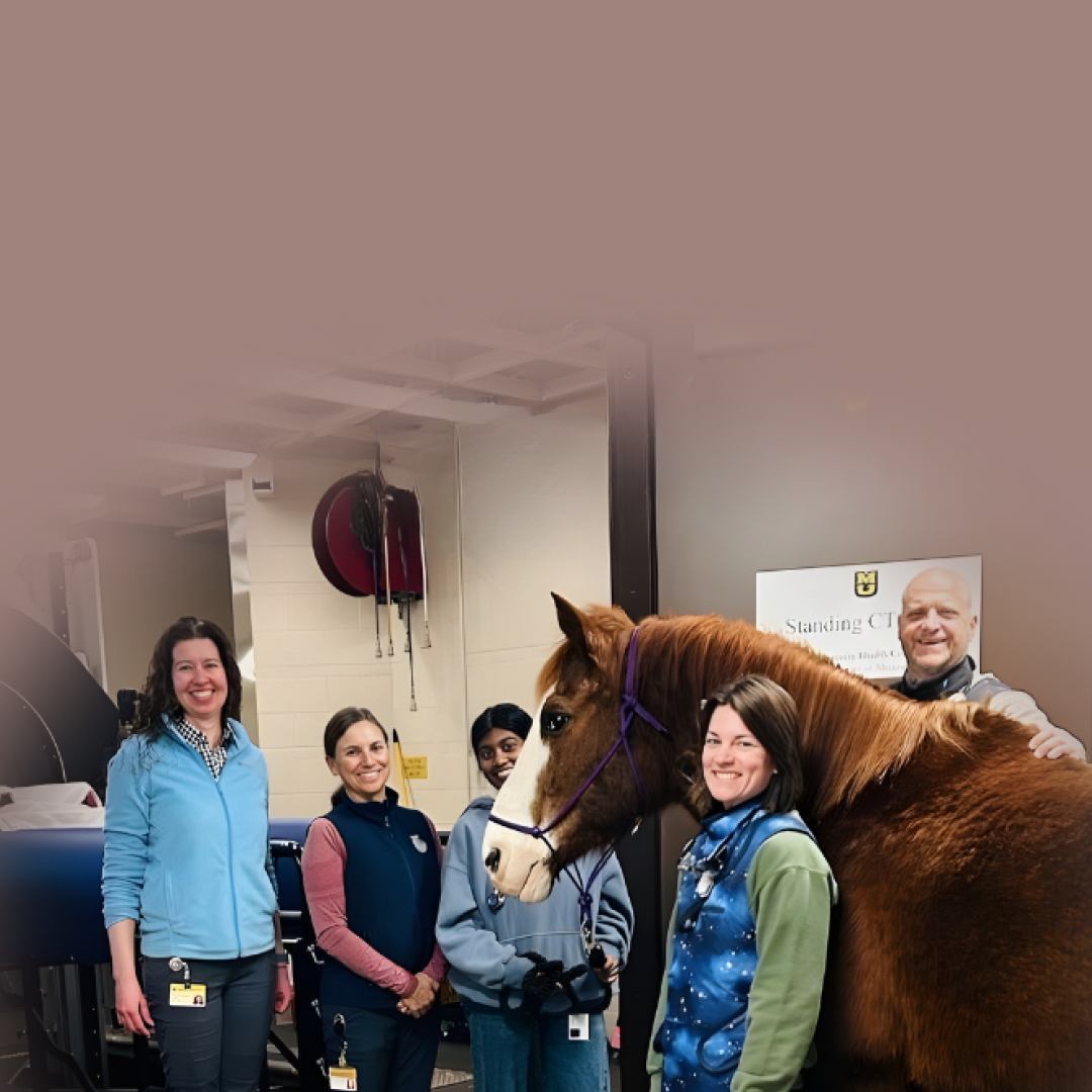
“Excited for our 500th Standing CT exam. In two and a half years, this diagnostic tool has revolutionized our equine practice. I see lesions that are not visible or difficult to interpret on radiographs almost every day”
Read more →
Megan J. McCracken
DVM, MS, DACVS-LA, Assistant Teaching Professor Equine Surgery, Missouri University Veterinary Health Center
Read More
Dr. Jack Caldwell
“In private practice, standing CT is practical and well tolerated by the horse. It requires less time than MRI, allows us to image a larger area of interest, and is often more cost effective. We’re identifying pathology beyond what ultrasound and radiographs”
Read more →
Dr. Jack Caldwell
DVM, Virginia Equine Imaging
Read More
Dr. Paul Wan
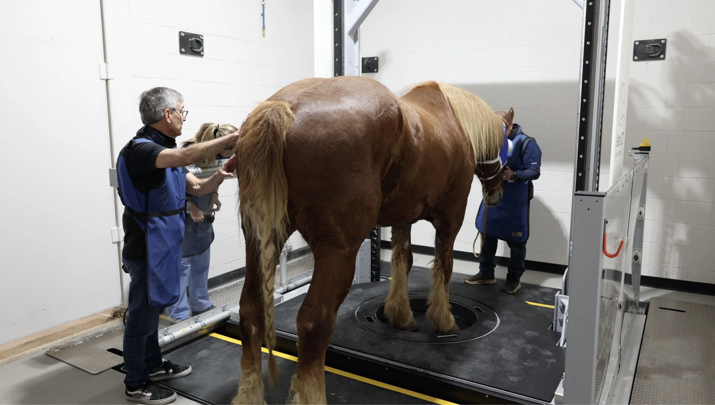
“The Equina® is one of the most innovative, advanced imaging devices available for veterinary medicine. We can scan front or back limb pairs, or the head/neck of a standing horse under light sedation, allowing the patient to be in and out in just minutes with minimal stress and risk.”
Read more →
Dr. Paul Wan
DVM, DACVS, CERT. VBM, SoCal Equine Hospital
Read More