

Shannon Vesledahl
B.S, CVT at UMN-Leatherdale Equine Center
“The Asto Equina isn't just a tool; it's transformed how we deliver care and diagnose issues in large animals, and I'm proud to be part of this advancement in veterinary medicine."

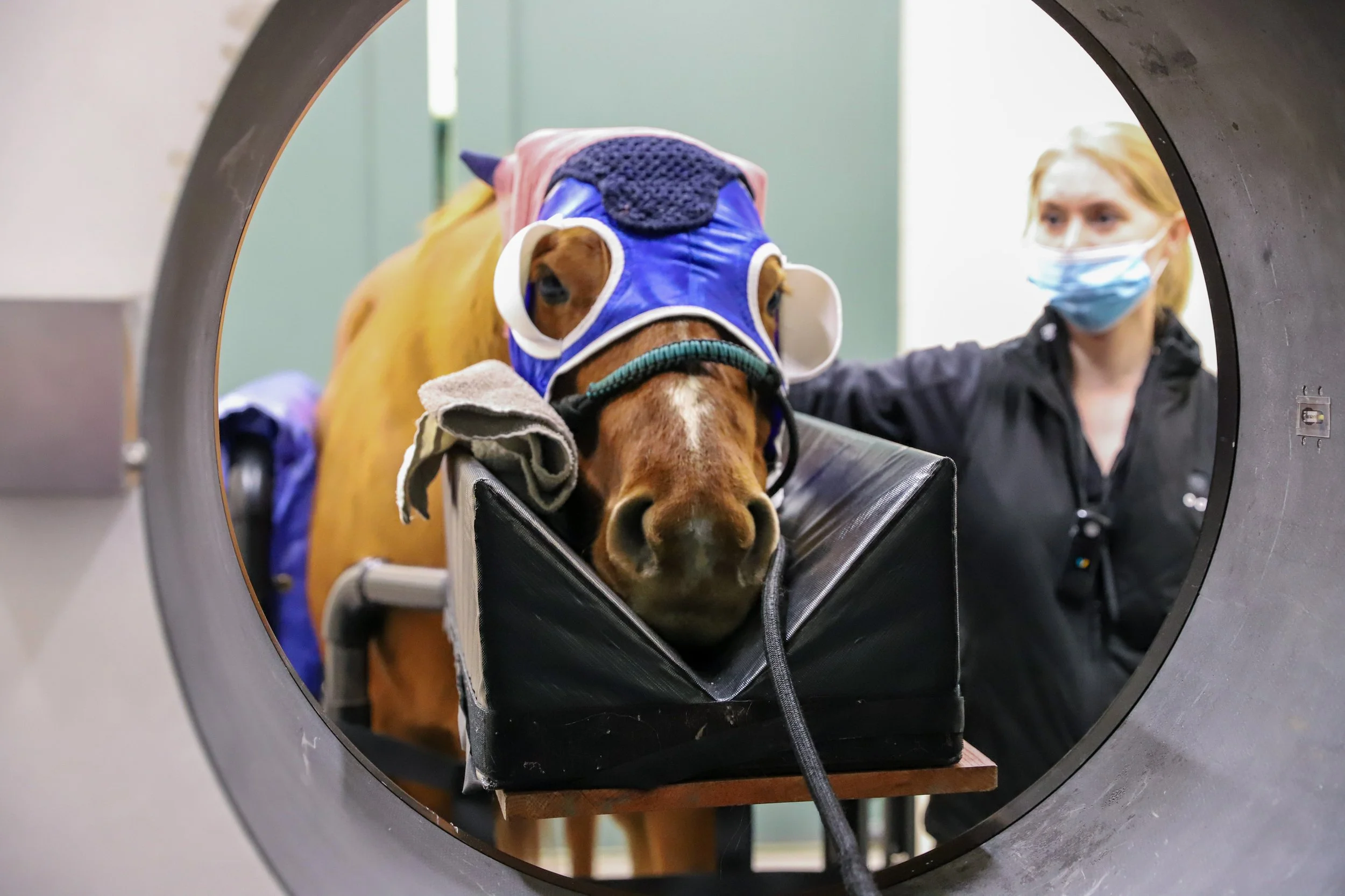
"As a surgery technician at the University of Minnesota Leatherdale Equine Center for over nine years, I've seen firsthand the evolution in diagnostic capabilities with the Asto Equina CT. Before its introduction in 2019, our diagnostic process often involved lengthy procedures and multiple sedations for horses. With the Asto Equina, we've revolutionized our approach. We can now achieve quicker and more accurate diagnoses with minimal sedation, significantly reducing the risk of complications like colic and saving owners money. Our efficiency has soared; what used to take days now takes minutes. We've uncovered hidden conditions like temporalhyoidosteopathy and fractures that were previously undetectable with traditional methods. As technicians, the CT has empowered us to independently manage scans, enhancing our role in patient care while allowing our veterinarians to focus on more cases. The Asto Equina isn't just a tool; it's transformed how we deliver care and diagnose issues in large animals, and I'm proud to be part of this advancement in veterinary medicine." —Shannon Vesledahl, large animal technician at UMN Leatherdale Equine Center

Read Shannon’s full blog article below
Case Study: Mandibular Mass in a 2-Month-Old QH Foal
History & Physical Exam
Patient: 2-month-old Quarter Horse foal
Presenting Complaint: Mass on the left side of the jaw, present since birth
Previous Workup:
Biopsy taken by referring DVM two days before CT
Results were pending at the time of CT
Systemic Condition: Otherwise well

Diagnosis
Mass Location: Rostral aspect of the right mandible
Differential Diagnoses:
Atypical cystic lesion
Developmental mass (teratoma)
Odontogenic neoplasm (though rare at this age)
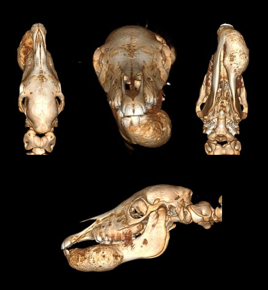
Imaging Findings:
Hyperattenuated areas similar to dental tissue
Mass extends to the symphysis but remains confined to the right mandible
Treatment & Outcome:
Final Diagnosis: Mandibular ossifying fibroma (confirmed via biopsy after CT)
Owner’s Decision: Chose to take the foal home for euthanasia