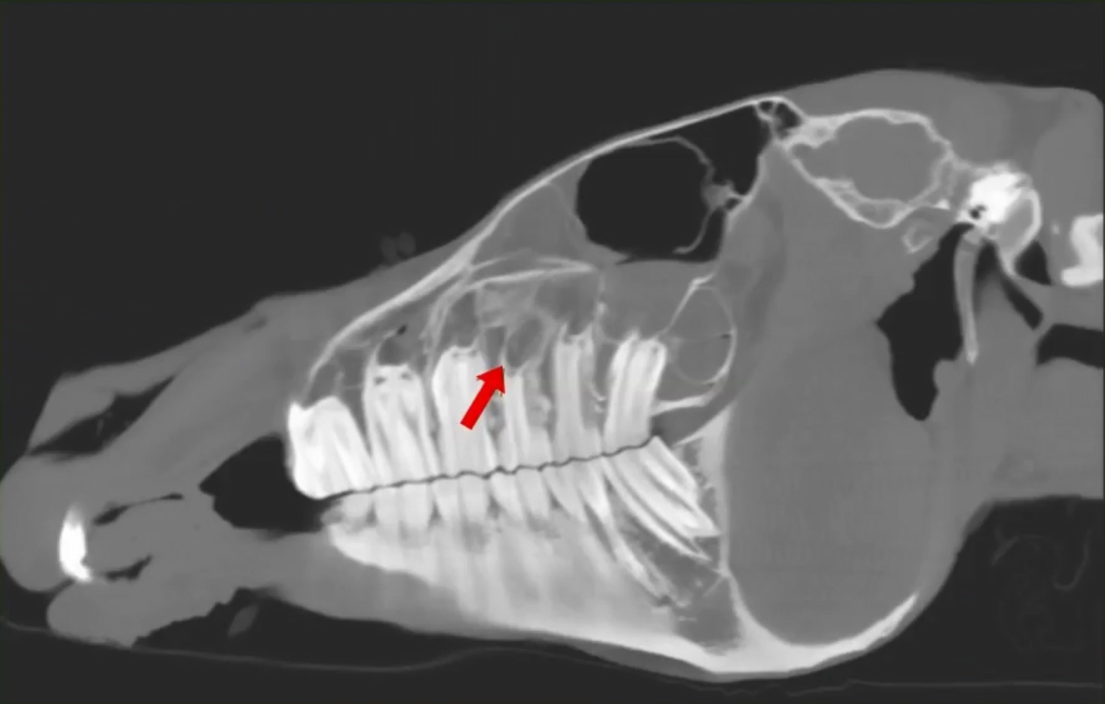
A 10-year-old TB mare was referred for evaluation of persistent right‑sided purulent nasal discharge that failed to respond to antibiotics. Despite significant nasal drainage, upper airway endoscopy was unremarkable, leaving the underlying cause unclear.
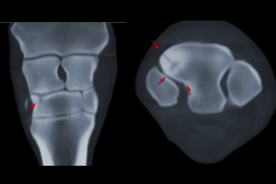
Traditional radiographs (including a skyline view) showed subtle changes but made the fracture difficult to visualize clearly. CT imaging provided a dramatic improvement in diagnostic clarity. The scan revealed a distinct fracture line, surrounding sclerosis, and areas of demineralization features that were not easily discernible on radiographs.
This case highlights the diagnostic power of CT with intra‑articular contrast for accurately assessing distal limb pathology, especially when MRI findings are equivocal or incomplete. CT provided a definitive understanding of the extent of cartilage loss and joint disease, ultimately guiding appropriate case management.
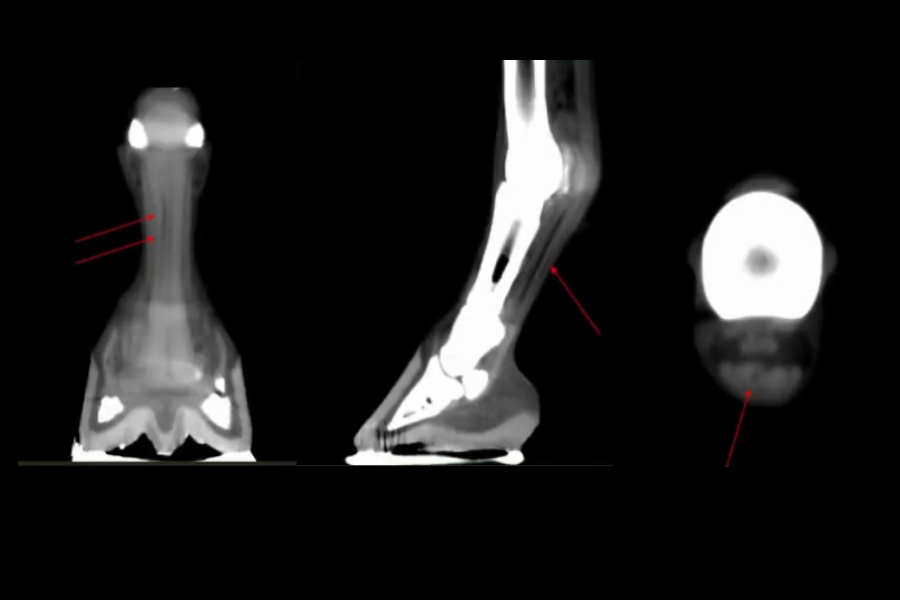
CT imaging revealed a moderately progressive DDFT tear now extending from mid‑P1 to the distal navicular bone, including a new full‑thickness parasagittal suprasesamoidean tear.
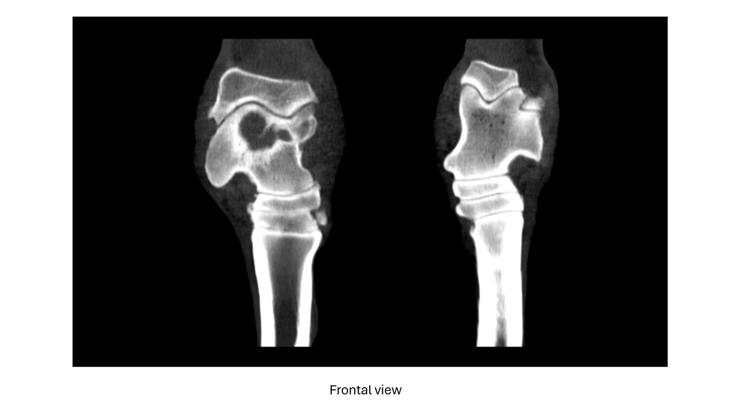
Advanced imaging revealed moderate tarsocrural joint osteoarthrosis and a large talar cyst with articular communication and an associated depression fracture, findings most consistent with a traumatic cyst.
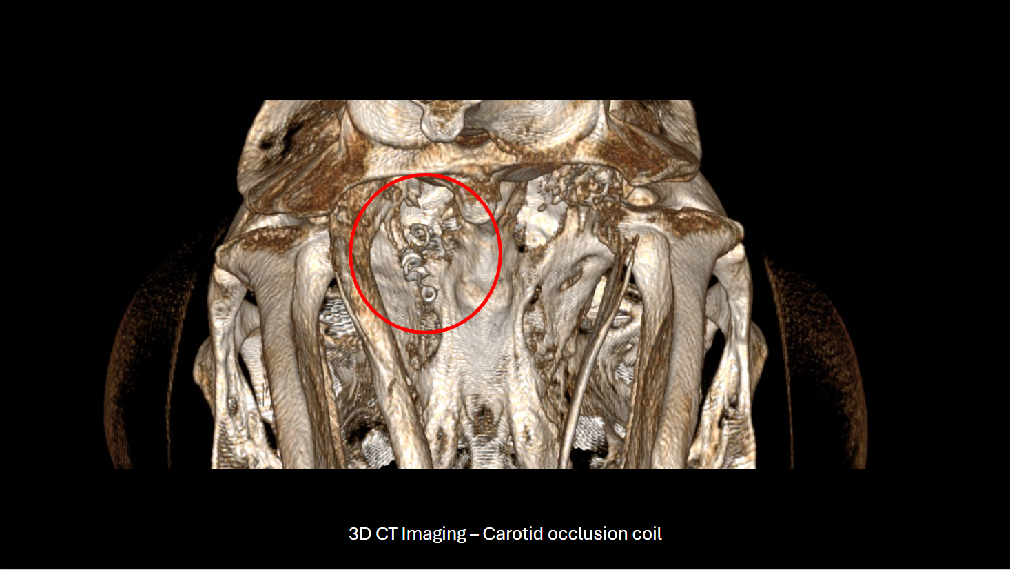
CT imaging demonstrated extensive occipital bone lysis and hyperostosis, regional infiltrative hypoattenuating soft tissue causing spinal cord compression, and marked thickening of the left stylohyoid bone and temporohyoid articulation
We're pleased to announce significant progress in deploying our technology to image the cervical spine. We were able to reach the cranial aspect of C7 at Virginia Equine Imaging on several standing patients.